. 2009 Jan;71(1):66-78.
doi: 10.1111/j.1365-2958.2008.06510.x. Epub 2008 Oct 28.

Novel Escherichia coli RF1 mutants with decreased translation termination activity and increased sensitivity to the cytotoxic effect of the bacterial toxins Kid and RelE

Affiliations

Novel Escherichia coli RF1 mutants with decreased translation termination activity and increased sensitivity to the cytotoxic effect of the bacterial toxins Kid and RelE

Elizabeth Diago-Navarro et al. Mol Microbiol. 2009 Jan.

Abstract

Novel mutations in prfA, the gene for the polypeptide release factor RF1 of Escherichia coli, were isolated using a positive genetic screen based on the parD (kis, kid) toxin-antitoxin system. This original approach allowed the direct selection of mutants with altered translational termination efficiency at UAG codons. The isolated prfA mutants displayed a approximately 10-fold decrease in UAG termination efficiency with no significant changes in RF1 stability in vivo. All three mutations, G121S, G301S and R303H, were situated close to the nonsense codon recognition site in RF1:ribosome complexes. The prfA mutants displayed increased sensitivity to the RelE toxin encoded by the relBE system of E. coli, thus providing in vivo support for the functional interaction between RF1 and RelE. The prfA mutants also showed increased sensitivity to the Kid toxin. Since this toxin can cleave RNA in a ribosome-independent manner, this result was not anticipated and provided first evidence for the involvement of RF1 in the pathway of Kid toxicity. The sensitivity of the prfA mutants to RelE and Kid was restored to normal levels upon overproduction of the wild-type RF1 protein. We discuss these results and their utility for the design of novel antibacterial strategies in the light of the recently reported structure of ribosome-bound RF1.
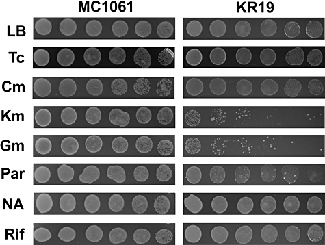

Figures

Fig. 1
Fig. 1
Complementation of a temperature-sensitive prfA mutation. MRA8 [prfA1(ts)] strain was transformed with the plasmids pELI01 (prfA121), pELI02 (prfAwt), pELI03 (prfA301) and pELI04 (prfA303). Expression of the prfA alleles in these plasmids was placed under the control of a promoter whose activity could be induced by adding arabinose into the medium. A. Uninduced cultures were grown at 42°C for up to 160 min. B. Same experiment but arabinose (1% final) was present in the medium to induce the expression of the different prfA alleles.
Fig. 2
Fig. 2
In vivo readthrough of the kis74 amber mutation in KR19 strain. Western blot analysis was performed using different cell extracts as described in Experimental procedures. The black arrow indicates full-length Kis protein. Lanes 1, 3 and 5, cell extracts from strain MC1061 bearing pFUS2, pELI05 (kis) and pELI06 (kis74) plasmids, respectively; lanes 2, 4 and 6, cell extracts from strain KR19 carrying pFUS2, pELI05 and pELI06 plasmids, respectively; lane 7, purified his-tagged Kis protein. The thick band at the bottom of the gel is a non-specific product present in whole-cell lysates.
Fig. 3
Fig. 3
Efficiency and levels of RF1 in prfA mutant strains in vivo. Termination was measured by competition with frameshifting at the shifty site present in the E. coli prfB gene (Poole et al., 1995). A. A region of 23 nucleotides from the prfB gene around the frameshift site was fused to the malE gene present in a plasmid under the control of the P-tac promoter. Frameshifting (+1) of the ribosome paused at the in-frame stop signal allows translation to continue into a sequence derived from the lacZ gene. The wild-type tetranucleotide stop signal TGAC was replaced by the tetranucleotides TAGA or TAGU. B. Expression of the MalE fusion products was induced in vivo in the parental strain MC1061 and each of the mutant strains KR4, KR17 and KR19 with IPTG for 2 h. The protein products of termination and frameshifting present in 3 or 6 μl of cell extract (see Experimental procedures) were separated on SDS gels and visualized by Western blotting using anti-MalE antibodies and 125I-labelled protein A. C. The amounts of RF1 in parental and prfA mutant strains were determined by quantitative Western blotting with rabbit anti-RF1 antibodies and 125I-labelled protein A.
Fig. 4
Fig. 4
Hypersensitivity of the prfA mutant strain KR19 to the Kid and RelE toxins and neutralization by cognate antitoxins and by prfAwt overexpression. Parental MC1061 and prfA mutant KR19 strains bearing the pNDM220 plasmid vector (lanes 1 and 2) or its derivatives encoding Kid (lanes 3 and 4) and RelE (lanes 5 and 6) toxins were grown exponentially to an A600 of 0.4 in LB medium. Serial dilutions (10−1 steps from left to right) of the different cultures were spotted in LB solidified medium. In (B) and (C), IPTG (100 μM) was included in the plates to induce the expression of the Kid and RelE toxins from the plasmids. In (A) and (D), the same experiment was performed but IPTG was omitted. Specificity controls were carried out by transforming the cells with plasmid pELI08 (prfAwt) (C and D) or with the plasmids encoding the respective antitoxins to Kid and RelE, namely plasmids pELI05 (kis) and pELI09 (relB) (E). The plates were incubated at 30°C and photographs were taken after 18 h incubation.
Fig. 5
Fig. 5
Inhibition of protein synthesis in vivo by the Kid and RelE toxins. Strains MC1061 (top) or KR19 (bottom) were transformed with plasmids as indicated: lane (1) plasmid vector pNDM220, lane (2) pSS100 (kid), lane (3) pELI07 (relE), lane (4) pSS100 (kid) and pELI05 (kis), lane (5) pELI07 (relE) and pELI09 (relB). When relevant, expression of the toxin and antitoxin genes was triggered by adding IPTG (100 μM) and arabinose (0.5%) to the medium respectively. The proteins synthesized at time 0 or 60 min after the induction of the toxin and antitoxin genes were fractionated by SDS-PAGE (12.5%) and identified by autoradiography as described in Experimental procedures.
Fig. 6
Fig. 6
Sensitivity assays of the KR19 (prfA301) and the parental strain MC1061 to different antibiotics. Parental MC1061 and prfA mutant KR19 strains were grown at 30°C to an A600 of 0.4 in LB medium. Serial dilutions (10−1 steps from left to right) of the different cultures were spotted in LB solidified medium (lane LB) as a positive control or in the same medium supplemented with the different antibiotics: tetracycline (Tc, 0.1 μg ml−1), chloramphenicol (Cm, 0.5 μg ml−1), kanamycin (Km, 1.0 μg ml−1), gentamicin (Gm, 0.5 μg ml−1), paromomycin (Par, 1.0 μg ml−1), nalidixic acid (NA, 1.0 μg ml−1) and rifampycin (Rif, 1.0 μg ml−1). Concentrations of the different antibiotics close to their minimal inhibitory concentrations were used. The plates were incubated at 30°C and photographs were taken after 18 h incubation.
Fig. 7
Fig. 7
Positions of the mutations in RF1 in relation to domains and sequence motifs of functional importance. The positions of residues G121, G301 and G303 for which mutants are found are shown space-filled in red (G121 and R203) or yellow (G301) on a model of the open form of Thermotoga maritima RF1 in ribbon representation (Vestergaard et al., 2005). The α5 helix, which has G121 at the tip, is shown in violet. The PxT motif implicated in recognition of the stop codon (Ito et al., 2000) is shown space-filled in pink, and residues T198 and Q185 interacting with the third stop codon nucleotide (Laurberg et al., 2008) are space-filled in light blue. The GGQ motif that interacts with the peptidyl transferase centre and triggers peptidyl-tRNA hydrolysis is shown space-filled in blue.
Fig. 8
Fig. 8
Ribosomal and mRNA components near to mutant residues in RF1. Components near residues G121, G301 and R303 in E. coli RF1 are shown, based on the 3.2 Å resolution crystal structure of Laurberg et al. (2008) of Thermus thermophilus RF1 bound to homologous 70S ribosomes in the presence of a short mRNA (5′-GGC AAG GAG GUA AAA A16U17G18U19A20A21 AAA AAA-3′; stop codon in bold; nucleotides 16–21 are shown in yellow) and P- and E-site bound tRNAMetf. The RF1 polypeptide is shown as a green ribbon (except for helix α5, in blue), with mutant residues space-filled (G121: red, G301: orange, R303: green); residues are numbered as in E. coli RF1. Residues in RF1 involved in stop codon recognition are also shown space-filled (P188 and T190: rose, Q185 and T198: blue). Nucleotides 1491–1498 in 16S rRNA are shown in grey, and ribosomal protein S12 is shown as a turquoise ribbon. See Table 3 for more information about near neighbours of the mutant residues in RF1.

Similar articles

Cited by

References

    1. Bachmann BJ. Pedigrees of some mutant strains of Escherichia coli K-12. Bacteriol Rev. 1972;36:525–557. - PMC - PubMed
    1. Baranov PV, Gesteland RF, Atkins JF. Release factor 2 frameshifting sites in different bacteria. EMBO Rep. 2002;3:373–377. - PMC - PubMed
    1. Bernard P, Couturier M. Cell killing by the F plasmid CcdB protein involves poisoning of DNA–topoisomerase II complexes. J Mol Biol. 1992;226:735–745. - PubMed
    1. Bravo A, Ortega S, de Torrontegui G, Diaz R. Killing of Escherichia coli cells modulated by components of the stability system ParD of plasmid R1. Mol Gen Genet. 1988;215:146–151. - PubMed
    1. Chopra I, Roberts M. Tetracycline antibiotics: mode of action, applications, molecular biology, and epidemiology of bacterial resistance. Microbiol Mol Biol Rev. 2001;65:232–260. - PMC - PubMed

Publication types

MeSH terms

LinkOut - more resources