. 2009 Feb;83(3):1402-15.
doi: 10.1128/JVI.01138-08. Epub 2008 Nov 19.

SOCS-1 mimetics protect mice against lethal poxvirus infection: identification of a novel endogenous antiviral system

Affiliations

SOCS-1 mimetics protect mice against lethal poxvirus infection: identification of a novel endogenous antiviral system

Chulbul M Ahmed et al. J Virol. 2009 Feb.

Abstract

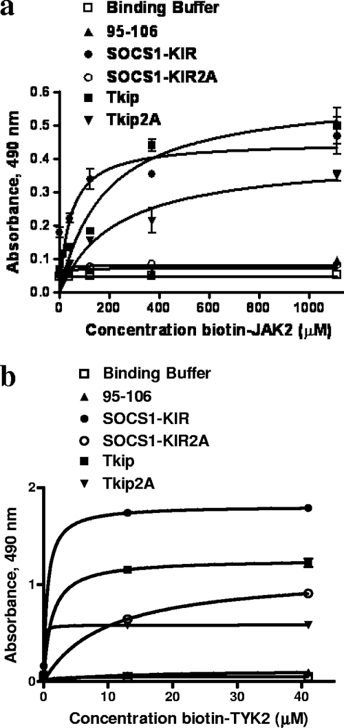
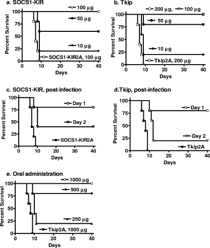
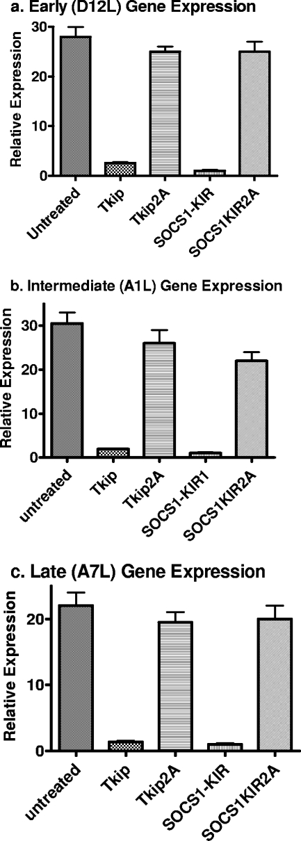
The suppressor of cytokine signaling 1 (SOCS-1) protein modulates cytokine signaling by binding to and inhibiting the function of Janus kinases (JAKs), ErbB, and other tyrosine kinases. We have developed a small tyrosine kinase inhibitor peptide (Tkip) that binds to the autophosphorylation site of tyrosine kinases and inhibits activation of STAT transcription factors. We have also shown that a peptide corresponding to the kinase-inhibitory region of SOCS-1, SOCS1-KIR, similarly interacts with the activation loop of JAK2 and blocks STAT activation. Poxviruses activate cellular tyrosine kinases, such as ErbB-1 and JAK2, in the infection of cells. We used the pathogenesis of vaccinia virus in C57BL/6 mice to determine the ability of the SOCS-1 mimetics to protect mice against lethal vaccinia virus infection. Injection of mice intraperitoneally with Tkip or SOCS1-KIR containing a palmitate for cell penetration, before and at the time of intranasal challenge with 2 x 10(6) PFU of vaccinia virus, resulted in complete protection at 100 microg. Initiation of treatment 1 day postinfection resulted in 80% survival. Administration of SOCS-1 mimetics by the oral route also protected mice against lethal effects of the virus. Both SOCS1-KIR and Tkip inhibited vaccinia virus transcription and replication at early and possibly later stages of infection. Vaccinia virus-induced phosphorylation of ErbB-1 and JAK2 was inhibited by the mimetics. Protected mice mounted a strong humoral and cellular response to vaccinia virus. The use of SOCS-1 mimetics in the treatment of poxvirus infections reveals an endogenous regulatory system that previously was not known to have an antiviral function.


Figures

FIG. 1.
FIG. 1.
Hydropathic profiles of the JAK kinase JAK2 and TYK2 autophosphorylation sites and sequence alignment. (a) Hydropathic profiles of murine JAK2 and TYK2 as determined by the Kyte-Doolittle hydropathic plot (18) with a window of three amino acids. The two JAKs have similar, but not identical, profiles, suggesting similar structures. (b) Alignment of the amino acid sequences of the autophosphorylation sites of murine JAK kinases JAK2 and TYK2.
FIG. 2.
FIG. 2.
SOCS1-KIR and Tkip bind to the autophosphorylation site peptides of JAK2 and TYK2. (a) SOCS1-KIR binds to JAK2 autophosphorylation site peptide JAK2(1001-1013). SOCS1-KIR, SOCS1-KIR2A (alanines substituted for phenylalanines at positions 56 and 59 of SOCS1-KIR), Tkip, Tkip2A, and a control peptide were immobilized in 96-well plates at 3 μg/well. Following blocking, various concentrations of biotinylated JAK2(1001-1013) peptide were added and the plates were incubated for 1 h. Bound biotinylated JAK2(1001-1013) was detected with an avidin-neutravidin-HRP conjugate. Absorbance was measured with a standard plate reader. There were statistically significant differences between SOCS1-KIR and SOCS1-KIR2A (P < 0.001) and between Tkip and Tkip2A (P < 0.001), as determined by the Mann-Whitney signed-rank test. (b) SOCS1-KIR and Tkip bind in a dose-dependent manner to biotinylated TYK2 autophosphorylation site peptide. There were statistically significant differences between SOCS1-KIR and SOCS1-KIR2A (P < 0.001) and between Tkip and Tkip2A (P < 0.001), as determined by the Mann-Whitney signed-rank test. All of the binding assays were carried out in triplicate, and the data are representative of at least three independent experiments. Where there are no error bars, the values were essentially identical.
FIG. 3.
FIG. 3.
SOCS1-KIR binding to autophosphorylation site peptides JAK2 and TYK2 as determined by an antibody ELISA. (a) SOCS1-KIR antibody is specific for the SOCS1-KIR peptide. SOCS1-KIR, SOCS1-KIR2A, and a control peptide were immobilized on a 96-well plate at 3 μg/well. Following blocking, various dilutions of SOCS1-KIR antibody were added and the plate was incubated for 1 h. Bound antibody was detected with a goat anti-rabbit IgG-HRP conjugate, followed by the addition of OPD substrate and 2N H2SO4. Absorbance was measured with a standard plate reader. (b) SOCS1-KIR binds to JAK2 and TYK2 autophosphorylation site peptides. JAK2, TYK2, and a control peptide were immobilized in a 96-well plate at 3 μg/well. Following blocking, various concentrations of SOCS1-KIR were added and the plate was incubated for 1 h. SOCS1-KIR antibody was added for 1 h of incubation, and bound SOCS1-KIR was detected as described for panel a.
FIG. 4.
FIG. 4.
Kinase inhibition patterns of SOCS1-KIR and Tkip in STAT1, STAT2, and STAT3 phosphorylation. (a) SOCS1-KIR and Tkip inhibit IFN-γ-induced STAT1 activation in L929 cells. L929 cells were seeded onto six-well plates at 1 × 10⁶ cells/well and pretreated with medium alone, lipo-Tkip, lipo-SOCS1-KIR, or a control peptide for 2 h at 37°C. Following 2 h of incubation in the presence or absence of IFN-γ (4,000 U/ml), the cells were washed and lysed. Whole-cell extracts were resolved by 12% SDS-PAGE, transferred to nitrocellulose membrane, and examined with pSTAT1 (pY701) antibody. The membrane was stripped and reprobed with STAT1 antibody. (b) SOCS1-KIR and Tkip inhibit IFN-τ-induced STAT1 and STAT3 activation in L929 cells and STAT2 activation in WISH cells. WISH cells were used here because of the availability of reagents. L929 cells were seeded into six-well plates at 1 × 10⁶/well and pretreated with medium alone, lipo-Tkip, lipo-SOCS1-KIR, or a control peptide for 2 h at 37°C. WISH cells were seeded onto six-well plates at 2.5 × 10⁵/well and pretreated with medium alone, lipo-Tkip, lipo-SOCS1-KIR, or a control peptide for 2 h at 37°C. Following 2 h of incubation in the presence or absence of IFN-τ (10,000 U/ml), the cells were washed and lysed. Whole-cell extracts were resolved by 12% SDS-PAGE, transferred to nitrocellulose membrane, and examined with antibodies to pSTAT1 (pY701), pSTAT2 (pY690), or pSTAT3 (pY705). The membrane was stripped and reprobed with STAT1, STAT2, or STAT3 antibody. (c) SOCS1-KIR2A and Tkip2A do not inhibit IFN-τ-induced STAT1 activation in L929 cells. L929 cells were seeded onto six-well plates at 1 × 10⁶/well and pretreated with medium alone, lipo-SOCS1-KIR, lipo-SOCS1-KIR2A, lipo-Tkip, lipo-Tkip2A, or a control peptide for 2 h at 37°C. Following 2 h of incubation in the presence or absence of IFN-τ (10,000 U/ml), the cells were washed and lysed. Whole-cell extracts were resolved by 12% SDS-PAGE, transferred to nitrocellulose membrane, and examined with pSTAT1 (pY701) antibody. The membrane was stripped and reprobed with STAT1 antibody.
FIG. 5.
FIG. 5.
SOCS1-KIR and Tkip protect mice against intranasal challenge with vaccinia virus. (a) SOCS1-KIR. Mice (C57BL/6, n = 5 per group in all mouse experiments) were pretreated i.p. on days −2, −1, and 0 with 100 μg (○), 50 μg (▪), or 10 μg (▾) of lipo-SOCS1-KIR peptide or 100 μg (□) of control peptide lipo-SOCS1-KIR2A. On day 0, vaccinia virus (2 × 10⁶ PFU) was administered intranasally. Survival of mice was followed for 40 days. The significance of differences between different treatments was measured by the log rank survival method, which gave P values of 0.002, 0.002, and 0.005 for the administration of 100, 50, and 10 μg of lipo-SOCS1-KIR versus the control peptide, respectively. (b) Tkip. Mice were pretreated on days −2, −1, and 0 with 200 μg (○), 100 μg (▿), 50 μg (▪), or 10 μg (•) of lipo-Tkip or 200 μg of the lipo-Tkip2A control (□). Infection with vaccinia virus was similar to that in panel a. Postinfection treatment with lipo-SOCS1-KIR or lipo-Tkip provides partial protection against vaccinia virus infection. (c) Postinfection lipo-SOCS1-KIR treatment. Mice (n = 5) were infected intranasally with 2 × 10⁶ PFU of vaccinia virus on day 0. Starting at day 1 (○) or 2 (▿) after infection, mice were treated i.p. with 200 μg of lipo-SOCS1-KIR for 3 consecutive days. Control peptide lipo-SOCS1-KIR2A (▪) at 200 μg was administered on days −2, −1, and 0. Survival was followed for 40 days. P values for the significance of differences between lipo-SOCS1-KIR treatment on days 1 and 2 versus the control peptide lipo-SOCS1-KIR2A were 0.002 and 0.005, respectively. (d) Postinfection lipo-Tkip treatment. Mice were infected with vaccinia virus in a manner similar to that described for panel a. Starting at day 1 (○) or 2 (▿) after infection, mice were treated i.p. with 200 μg of lipo-Tkip for 3 consecutive days. Control peptide lipo-Tkip2A (▪) at 200 μg was administered on days −2, −1, and 0. Survival was followed for 40 days. P values for the significance of differences between lipo-Tkip treatment on days 1 and 2 versus the control lipo-Tkip2A were 0.002 and 0.005, respectively. (e) Oral treatment with lipo-Tkip protects mice against intranasal challenge with vaccinia virus. Mice (n = 5) were given 1,000 μg (○), 500 μg (•), or 250 μg (▴) of lipo-Tkip on days −2, −1, and 0 by the oral route. One thousand micrograms of a control peptide, lipo-Tkip2A (▪), was given orally on the same days. On day 0, 2 × 10⁶ PFU of vaccinia virus was administered intranasally. Survival of mice was followed for 40 days. P values for the significance of differences between 1,000, 500, and 250 μg of lipo-Tkip and the control peptide were 0.002, 0.008, and 0.008, respectively.
FIG. 6.
FIG. 6.
One-step growth curve of inhibition of vaccinia virus replication by SOCS1-KIR and Tkip. BSC-40 cells grown to confluence were left untreated or treated with lipo-SOCS1-KIR, lipo-Tkip, or their alanine substitution-containing mutant forms at 50 μM for 1 h. For a positive control, an IFN-γ mimetic spanning residues 95 to 132 (designated 95-132) that was previously shown to inhibit vaccinia virus replication (1, 3) was used. Cells were then infected with vaccinia virus at an MOI of 5 for 1 h, after which the cells were washed and incubated in the presence of the same concentrations of peptides for the indicated times. The cell extracts (a) and supernatants (b) obtained were titrated for the amounts of intracellular and extracellular virus, respectively. Note the difference of the scale on the y axis, indicating that there is less extracellular virus than intracellular virus.
FIG. 7.
FIG. 7.
SOCS1-KIR and Tkip inhibit vaccinia virus replication in a dose-dependent manner. BSC-40 cells were grown to confluence and left untreated or treated with the indicated amounts of lipo-SOCS1-KIR (a and b) or lipo-Tkip (c and d) for 1 h. Control peptides lipo-SOCS1-KIR2A (a and b) and lipo-Tkip2A (c and d) were used at 25 μM. Cells were next infected with vaccinia virus at an MOI of 5. After 1 h, the cells were washed and incubated in the presence of the same concentrations of peptides for 1 day. The supernatants and cell extracts obtained were titrated for the amounts of intracellular (a and c) and extracellular (b and d) virus, respectively.
FIG. 8.
FIG. 8.
Adaptive immune response in mice that recovered from vaccinia virus infection with lipo-Tkip treatment. (a) Survival of mice after a rechallenge with vaccinia virus. Naïve mice (▪; n = 5) and those that had recovered from vaccinia virus infection with lipo-Tkip treatment for 30 days (▾) were infected intranasally with 2 × 10⁶ PFU of vaccinia virus in 10 μl. Survival was followed for 50 days. The significance of the difference, as measured by log rank survival, was P = 0.0002 for the rechallenged group versus the naïve mice. (b) Cell-mediated immune response in mice that recovered from vaccinia virus infection with lipo-Tkip treatment. Splenocytes (105) obtained from naïve mice or mice that recovered (n = 3) 2 or 3 weeks after infection and lipo-Tkip treatment were incubated with UV-inactivated, purified vaccinia virus. Four days later, [3H]thymidine was added for 8 h of incubation and its incorporation was followed. The stimulation index is the incorporation into splenocytes cultured with test antigen divided by the incorporation into splenocytes cultured with medium alone. Averages and standard deviations are shown. (c) Vaccinia virus-specific response in CD4-depleted splenocytes by ELISPOT analysis. Splenocytes obtained from naïve mice or mice that recovered (n = 3) 2 or 3 weeks after infection with lipo-Tkip treatment were depleted of CD4 cells, and 105 cells thus obtained were incubated in microtiter plates previously coated with antibody to IFN-γ in the presence of vaccinia virus (MOI = 0.01). After 2 days of incubation, the spots (IFN-γ-secreting cells) per well were counted. The values shown are averages and standard deviations. (d and e) Presence of vaccinia virus-specific antibodies in mice that recovered from infection with lipo-Tkip treatment. Sera collected from naïve mice (n = 3) or those that recovered from vaccinia virus infection with lipo-Tkip treatment were collected in the weeks indicated. Sera were diluted as indicated and added to wells of microtiter plates coated with UV-inactivated vaccinia virus. After washing to remove nonspecific binding, secondary anti-mouse IgA (d) or IgG (e) antibody conjugated to HRP was added, followed by the addition of OPD substrate and absorption measurement. The values shown are averages and standard deviations. (f) Neutralizing antibodies in mice that recovered. Sera taken from naïve mice (n = 3) or those that recovered from vaccinia virus infection with lipo-Tkip treatment in the weeks indicated were diluted as shown, mixed with vaccinia virus (100 PFU), incubated for 1 h, and then added to BSC-40 cells. One hour later, regular growth medium was added to the cells and the mixture was incubated for 2 days. The reduction in the number of plaques is shown as a percentage and the standard deviation.
FIG. 9.
FIG. 9.
Vaccinia virus-induced phosphorylation of ErbB-1 and JAK2 is inhibited by Tkip. Extracts of BSC-40 cells left untreated or pretreated for 30 min with 20 μM lipo-Tkip or control peptide lipo-Tkip2A and infected with vaccinia virus (MOI = 0.1) for 1 or 5 min were harvested and immunoprecipitated with phosphotyrosine antibody. Phosphotyrosine proteins were electrophoresed and probed with antibodies to ErbB-1 or JAK2.
FIG. 10.
FIG. 10.
Inhibition of vaccinia virus transcription by Tkip and SOCS1-KIR. BSC-40 cells mock treated or treated with lipo-Tkip, lipo-SOCS1-KIR, or alanine substitution-containing control peptides for 1 h were infected with vaccinia virus at an MOI of 5 for 1 h. The cells were washed and incubated in growth medium containing the same concentration of peptides for 18 h. RNA was extracted and used for cDNA synthesis, followed by quantitative PCR. The expression of early (D12L) (a), intermediate (AIL) (b), and late (A7L) (c) genes was compared with endogenous actin gene expression.
FIG. 11.
FIG. 11.
Effect of SOCS1-KIR and Tkip treatment at different times on the replication of vaccinia virus. BSC-40 cells were infected with vaccinia virus at an MOI of 5 for 1 h. Cells were washed and treated with 25 μM lipo-SOCS1-KIR, lipo-Tkip, or the control peptides at time zero and 1 h, 2 h, and 4 h after infection. Cells were allowed to grow for 24 h. Cell extracts and supernatants and were harvested and assayed for the virus in cell extracts (a to d) and supernatants (e to h).

References

    1. Ahmed, C. M., M. A. Burkhart, M. G. Mujtaba, and H. M. Johnson. 2005. Peptide mimetics of gamma interferon possess antiviral properties against vaccinia and other viruses in the presence of poxvirus B8R protein. J. Virol. 795632-5639. - PMC - PubMed
    1. Ahmed, C. M. I., and H. M. Johnson. 2006. IFN-γ and its receptor subunit IFNGR1 are recruited to the IFN-γ-activated sequence element at the promoter site of IFN-γ-activated genes: evidence of transactivational activity in IFNGR1. J. Immunol. 177315-321. - PubMed
    1. Ahmed, C. M., J. P. Martin, and H. M. Johnson. 2007. IFN mimetic as a therapeutic for lethal vaccinia virus infection: possible effects on innate and adaptive immune responses. J. Immunol. 1784576-4583. - PubMed
    1. Alexander, W. S., and D. J. Hilton. 2004. The role of suppressors of cytokine signaling (SOCS) proteins in regulation of immune response. Annu. Rev. Immunol. 22503-529. - PubMed
    1. Ausubel, F. M., R. Brent, R. E. Kingston, D. D. Moore, J. G. Seidman, A. Smith, and K. Struhl (ed.). 2002. Short protocols in molecular biology, p. 16-65. Wiley, New York, NY.

Publication types

Substances