. 2009 Feb;83(3):1201-15.
doi: 10.1128/JVI.01797-08. Epub 2008 Nov 19.

Vaccinia virus extracellular enveloped virion neutralization in vitro and protection in vivo depend on complement

Affiliations

Vaccinia virus extracellular enveloped virion neutralization in vitro and protection in vivo depend on complement

Mohammed Rafii-El-Idrissi Benhnia et al. J Virol. 2009 Feb.

Abstract

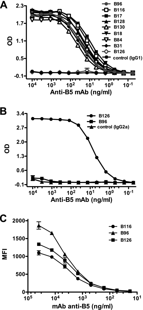
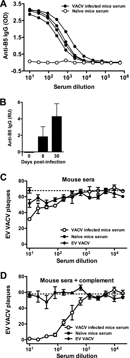
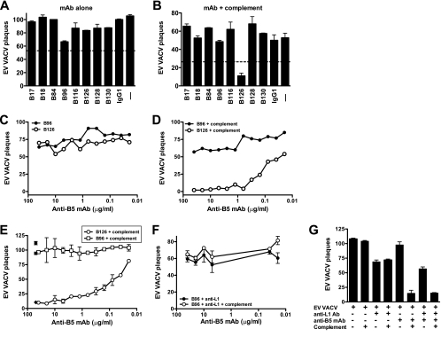
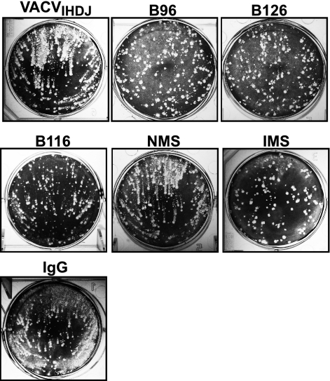
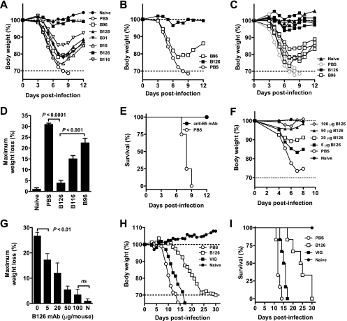
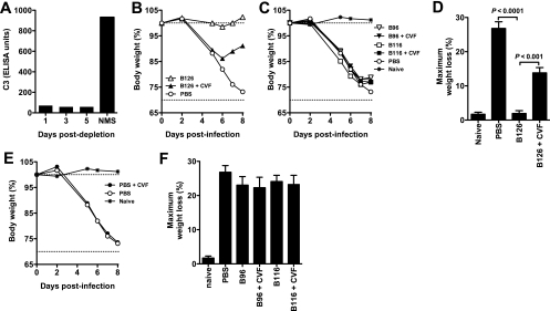
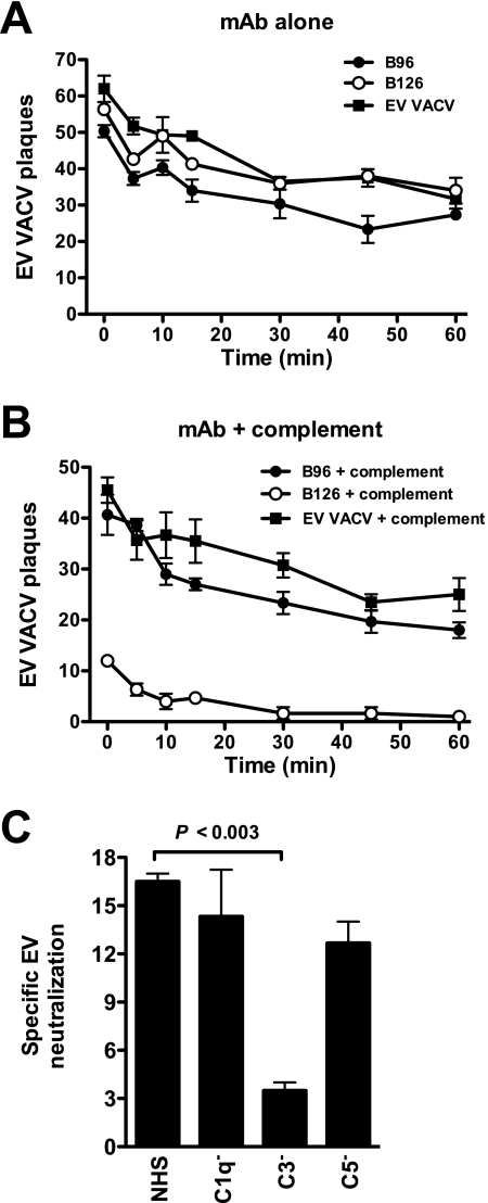
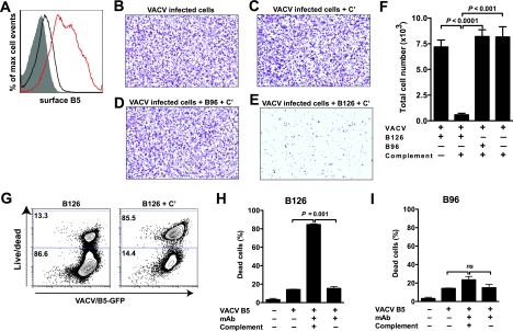
Antibody neutralization is an important component of protective immunity against vaccinia virus (VACV). Two distinct virion forms, mature virion and enveloped virion (MV and EV, respectively), possess separate functions and nonoverlapping immunological properties. In this study we examined the mechanics of EV neutralization, focusing on EV protein B5 (also called B5R). We show that neutralization of EV is predominantly complement dependent. From a panel of high-affinity anti-B5 monoclonal antibodies (MAbs), the only potent neutralizer in vitro (90% at 535 ng/ml) was an immunoglobulin G2a (IgG2a), and neutralization was complement mediated. This MAb was the most protective in vivo against lethal intranasal VACV challenge. Further studies demonstrated that in vivo depletion of complement caused a >50% loss of anti-B5 IgG2a protection, directly establishing the importance of complement for protection against the EV form. However, the mechanism of protection is not sterilizing immunity via elimination of the inoculum as the viral inoculum consisted of a purified MV form. The prevention of illness in vivo indicated rapid control of infection. We further demonstrate that antibody-mediated killing of VACV-infected cells expressing surface B5 is a second protective mechanism provided by complement-fixing anti-B5 IgG. Cell killing was very efficient, and this effector function was highly isotype specific. These results indicate that anti-B5 antibody-directed cell lysis via complement is a powerful mechanism for clearance of infected cells, keeping poxvirus-infected cells from being invisible to humoral immune responses. These findings highlight the importance of multiple mechanisms of antibody-mediated protection against VACV and point to key immunobiological differences between MVs and EVs that impact the outcome of infection.


Figures

FIG. 1.
FIG. 1.
Binding affinities of anti-B5 MAbs. Titration of anti-B5 MAbs using IgG1-specific (A) and IgG2a-specific (B) ELISAs. (C) Mean fluorescence intensity (MFI) of cell-based ELISA quantitating surface-bound anti-B5 MAbs to native B5 on VACV-infected cells. Serial dilution of each anti-B5 MAb was performed. Data are representative of three independent experiments. OD, optical density.
FIG. 2.
FIG. 2.
Anti-B5 IgG responses in VACV-infected mice. (A) B5 ELISA of serum from VACV-infected mice at day 8 postinfection. (B) Quantitation of anti-B5 IgG by protein microarray at days 8 and 30 postinfection. (C and D) VACV EV neutralizing antibody activity of the day 30 mouse serum described above in the absence (C) or presence (D) of complement. Plaque assay results are graphed. A dashed line indicates the plaque number of VACV EV alone (C) or in the presence of complement without antibody (D). Data are representative of two experiments. Error bars indicate SEM for each condition. OD, optical density; RU, relative units.
FIG. 3.
FIG. 3.
Complement-dependent MAb neutralization of EV. VACV EV neutralization is dependent on complement and anti-B5 monoclonal antibody isotype. (A and B) VACV EV neutralization activity of the full panel of MAbs against B5 at 10 μg/ml in the absence (A) or the presence (B) of complement. EV alone (− lane) and mouse anti-dinitrophenol control IgG1 plus EV (IgG1) were negative controls. (C and D) Titrated VACV EV neutralization activity of murine anti-B5 MAbs B126 (IgG2a) and B96 (IgG1) in the absence (C) or the presence (D) of complement. Data are represented as plaque numbers. The 50% and 90% neutralization titers of B126 (107 ng/ml and 535 ng/ml, respectively) were determined based on sigmoidal dose-response nonlinear regression. All data are representative of three or more experiments, each of which was done in the presence of anti-L1 antibody. Error bars indicate SEM in each condition. (E to G) Titrated VACV EV neutralization activity of murine anti-B5 MAbs B126 (IgG2a) and B96 (IgG1) in the absence or the presence of complement and with or without rabbit anti-L1 antibody. (E) In the absence of anti-L1 antibody, the neutralization activity of B126 plus complement or B96 plus complement was titrated. Control samples are shown at single dilutions: B126 alone (filled circle), B96 alone (filled square), and B126 with complement and rabbit anti-L1 antibody (filled diamond). (F) Titration of B96 plus anti-L1 antibody and B96 plus anti-L1 antibody and complement. (G) Quantitation of conditions described in panels E and F and additional controls, with anti-B5 MAb B126 at 10 μg/ml. Data in panels E to G are representative of three independent experiments. Error bars indicate SEM for each condition.
FIG. 4.
FIG. 4.
Comet tail plaque inhibition. All anti-B5 MAbs exhibited comet tail inhibition activity in vitro, independent of isotype. Vero E6 cells were infected with VACVIHDJ and then cultured in the absence of antibody (VACVIHDJ) or with anti-B5 MAbs (B96, B126, or B116), naïve mouse serum (NMS), immune mouse serum (IMS), or irrelevant MAb (IgG).
FIG. 5.
FIG. 5.
Protection against a lethal VACV infection. All anti-B5 MAbs exhibited some protective effect in vivo, and B126 IgG2a was highly protective. (A to E) Groups of BALB/c mice were inoculated i.p. with 100 μg of anti-B5 MAb (B18, B31, B96, B116, B126, or B128). Control mice received PBS. Naïve mice were unchallenged. One day later, mice were challenged intranasally with 5 × 10⁴ PFU of purified VACV. Body weight was tracked, and average weights were graphed (A). Dotted lines indicate starting body weight (upper) and terminal body weight (bottom). B126 and B96 group means are shown again for clarity (B), with all individual animal curves are shown (C), and weight loss nadirs were calculated for specific experimental groups (D). B126-treated mice exhibited no significant weight loss compared to uninfected mice (P ≫ 0.05) and were significantly better protected than mice treated with any other MAb (P < 0.001; n = 4/group). Panel E shows survival after intranasal challenge. All MAb-treated mice were protected from death compared to mock-treated mice (P < 0.01, for each individual MAb versus PBS). One of four independent experiments is shown. (F and G) Dose titrations of MAb B126 were tested against intranasal challenge with VACV as described above. Weight loss curves for each group (F) and percent maximum weight loss (G) were plotted. Five micrograms of anti-B5 MAb B126 completely prevented mortality, and mice exhibited significantly ameliorated weight loss versus no MAb (P < 0.01; n = 6/group). A 100-μg dose provided excellent protection, with virtually no weight loss observed (P = 0.5; ns). One of five independent experiments is shown. (H and I) SCID mice were inoculated with 100 μg of anti-B5 MAb B126 or 1.25 mg of VIG. Body weight was tracked after intravenous infection with 1 × 10³ PFU VACV. Mean percentage weight loss (H) and survival curves (I) were plotted for each experimental group. Anti-B5 MAb B126 protected substantially better than VIG, as determined by weight loss (P < 0.0005 versus no treatment; P < 0.0014 versus VIG; n = 6/group) and survival (P < 0.001 versus no treatment; P < 0.01 versus VIG). Bar graph error bars indicate SEM for each condition.
FIG. 6.
FIG. 6.
Anti-B5 protection in vivo is complement dependent. Complement depletion in vivo abrogated the majority of anti-B5 protection against VACV. (A) Complement C3 levels in CVF-treated mice at days 1 to 5 after treatment. Serum from mice prior to treatment (naïve mouse serum [NMS]) was used as control. (B to D) Complement-depleted (+CVF) or nondepleted mice were treated with 100 μg of anti-B5 MAb (B96, B116, or B126) or PBS at day −1 and challenged intranasally with 5 × 10⁴ PFU of purified VACV at day 0. Mean weight loss kinetics in each group (B and C), and maximum weight loss (weight nadir) (D) are shown. As shown in panel B, VACV-infected mice treated with B126 were fully protected compared with untreated infected mice (P < 0.0001), but complement-depleted mice had a >50% specific loss in B126 MAb protection. As shown in panel C, B96 and B116 provided minimal protection against disease, and neither B96 nor B116 was affected by CVF treatment. Abrogation of anti-B5 B126 protection by complement-depletion was highly statistically significant (P < 0.0001, for B126 plus CVF versus B126 in complement-sufficient recipients B126), while there were no significant effect of complement depletion on any other experimental group (P ≫ 0.05; nonsignificant) (D). Complement depletion in vivo (CVF treatment) did not affect disease in untreated mice (E) or the modest activities of B96 or B116 (F). One of three independent experiments is shown. Error bars indicate SEM for each group.
FIG. 7.
FIG. 7.
Complement kinetics and components. (A and B) Kinetics of VACV EV neutralization activity in vitro of murine anti-B5 MAbs (B126 and B96) at 10 μg/ml in the absence or the presence of rabbit complement. Data are shown as plaque numbers. One of three independent experiments is shown. VACV EV is fully neutralized by anti-B5 MAb B126 within 5 min of the addition of complement. (C) VACV EV neutralization by the anti-B5 MAb and the addition of normal human serum (NHS) complement or C1q-, C3-, or C5-depleted serum. Specific VACV EV neutralization in the absence of C3 was statistically significant compared to the value for normal human serum (P < 0.003). One of three independent experiments is shown. Error bars indicate SEM for each group.
FIG. 8.
FIG. 8.
The purified VACV MV stock did not contain EV or B5 antigen. ELISA plates were coated with UV-inactivated VACV MV and then probed with serial dilutions of anti-B5 (B126) or anti-H3 (41) MAbs. Starting concentrations were 0.62 and 0.5 μg/ml, respectively. OD, optical density.
FIG. 9.
FIG. 9.
Complement and anti-B5 IgG2a cooperate to efficiently mediate destruction of VACV-infected cells. Anti-B5 antibodies are able to direct complement lysis of VACV-infected cells due to their surface expression of B5. (A) Cell monolayers (Vero E6) were infected with VACV (MOI of 5), and surface expression of B5 was tested at 4 h (black line) and 8 h (red line) after infection by surface-staining infected cells with anti-B5 MAb and performing flow cytometry. Uninfected cells, negative control (filled curve). (B to F) Anti-B5 directed complement lysis of infected cells. Virus-infected Vero E6 monolayer cells (crystal violet stained) at a magnification of ×40. VACV-infected cells were treated with medium (B) or complement (+ C′) (C to E) in the absence (C) or presence of anti-B5 IgG1 MAb B96 (D) or IgG2a MAb B126 (E). VACV-infected cells were completely and specifically destroyed by anti-B5 IgG2a and complement. (F) Quantitation of live cell numbers. Destruction of VACV-infected cells was highly statistically significant in the presence of anti-B5 MAb B126 and complement versus B126 alone (P < 0.0001), complement alone (P < 0.001), or B96 plus complement (P < 0.0001). No killing was observed for IgG1 B96 in the absence or presence of complement (P ≫ 0.05; nonsignificant). (G to I) Anti-B5 directed killing of virus-infected cells was assessed in a separate series of experiments using flow cytometric assays. Cells were infected with VACV-B5-GFP and treated with anti-B5 MAb B126 in the absence (left panel) or presence (right panel) of complement, and cell killing was determined by measuring the uptake of a viability dye (live/dead) by damaged cells (G). Killed infected cells exhibit intense live/dead fluorescence staining (y axis). Infected cells were identified by B5-GFP expression (x axis). Complement-mediated cell killing by anti-B5 IgG2a B126 was effective and highly statistically significant (P = 0.001 versus B126 alone or complement alone) (H). When IgG1 MAb B96 in the absence or presence of complement was used, no statistically significant (ns) differences were observed (I). Error bars indicate SEM for each group. One of three independent experiments is shown in panels A to F. One of two independent experiments is shown in panels G to I.

References

    1. Aldaz-Carroll, L., J. C. Whitbeck, M. Ponce de Leon, H. Lou, L. Hirao, S. N. Isaacs, B. Moss, R. J. Eisenberg, and G. H. Cohen. 2005. Epitope-mapping studies define two major neutralization sites on the vaccinia virus extracellular enveloped virus glycoprotein B5R. J. Virol. 796260-6271. - PMC - PubMed
    1. Aldaz-Carroll, L., J. C. Whitbeck, M. Ponce de Leon, H. Lou, L. K. Pannell, J. Lebowitz, C. Fogg, C. L. White, B. Moss, G. H. Cohen, and R. J. Eisenberg. 2005. Physical and immunological characterization of a recombinant secreted form of the membrane protein encoded by the vaccinia virus L1R gene. Virology 34159-71. - PubMed
    1. Aldaz-Carroll, L., Y. Xiao, J. C. Whitbeck, M. Ponce de Leon, H. Lou, M. Kim, J. Yu, E. L. Reinherz, S. N. Isaacs, R. J. Eisenberg, and G. H. Cohen. 2007. Major neutralizing sites on vaccinia virus glycoprotein B5 are exposed differently on variola virus ortholog B6. J. Virol. 818131-8139. - PMC - PubMed
    1. Amanna, I. J., M. K. Slifka, and S. Crotty. 2006. Immunity and immunological memory following smallpox vaccination. Immunol. Rev. 211320-337. - PubMed
    1. Bell, E., M. Shamim, J. C. Whitbeck, G. Sfyroera, J. D. Lambris, and S. N. Isaacs. 2004. Antibodies against the extracellular enveloped virus B5R protein are mainly responsible for the EEV neutralizing capacity of vaccinia immune globulin. Virology 325425-431. - PubMed

Publication types

Substances

LinkOut - more resources