. 2009 Jan;20(1):68-77.
doi: 10.1681/ASN.2008010036. Epub 2008 Nov 19.

Imatinib suppresses cryoglobulinemia and secondary membranoproliferative glomerulonephritis

Affiliations

Imatinib suppresses cryoglobulinemia and secondary membranoproliferative glomerulonephritis

Masayuki Iyoda et al. J Am Soc Nephrol. 2009 Jan.

Abstract

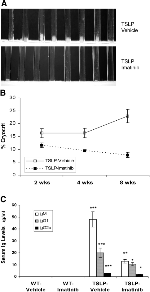
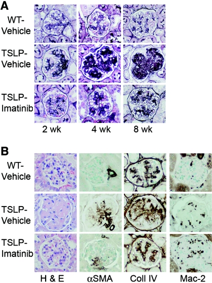
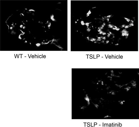
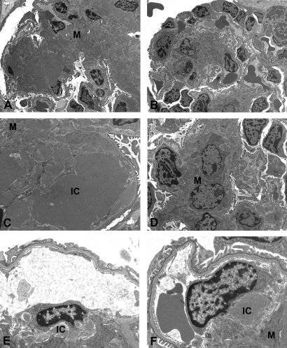
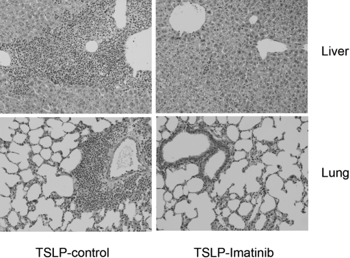
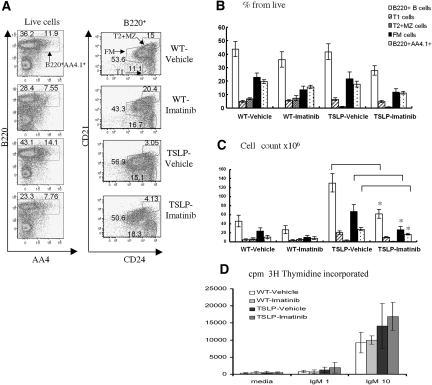
Imatinib is a receptor tyrosine kinase inhibitor that blocks the activity of c-Abl, c-Kit, and PDGF receptors. We tested the protective effects of imatinib in thymic stromal lymphopoietin transgenic mice, a model of cryoglobulinemia and associated membranoproliferative glomerulonephritis (MPGN), in which some glomerular manifestations likely result from PDGF receptor activation. Surprising, administration of imatinib beginning at weaning suppressed production of cryoglobulin, attenuating both the renal injury and systemic features of cryoglobulinemia. Flow cytometry suggested that inhibition of B cell development in the bone marrow likely caused the reduction in cryoglobulin production. In addition, administration of imatinib to thymic stromal lymphopoietin transgenic mice with established MPGN also diminished cryoglobulin production and reversed the renal and systemic lesions. These data suggest that treatment with imatinib may be a novel therapeutic approach for cryoglobulinemia and MPGN in humans.


Figures

Figure 1.
Figure 1.
Imatinib decreases cryoglobulin levels and serum Ig repertoire in TSLP-Tg mice. (A) The sera from vehicle-treated TSLP-Tg mice demonstrate visible cryoprecipitates but are dramatically reduced after 4 wk of treatment with imatinib. (B) Compared with vehicle-treated TSLP-Tg mice, a significant decrease of cryocrit is observed at each time point. Data are means ± SEM; n = 8 mice per group. Mann-Whitney test: *P < 0.05, **P < 0.01, ***P < 0.001: vehicle-treated TSLP-Tg mice versus imatinib-treated TSLP-Tg mice. (C) A significant decrease of serum Ig repertoire is seen after 4 wk of treatment with imatinib in TSLP-Tg mice. Data are means ± SEM; n = 6 to 8 mice per group. ANOVA, followed by Tukey-Kramer test: *P < 0.05, **P < 0.01, ***P < 0.001: vehicle-treated TSLP-Tg mice versus imatinib-treated TSLP-Tg mice; †P < 0.001: vehicle-treated WT mice versus vehicle-treated TSLP-Tg mice.
Figure 2.
Figure 2.
Imatinib attenuates glomerulonephritis in TSLP-Tg mice. (A) TSLP-Tg mice treated with vehicle (middle) develop progressive accumulation of matrix and deposits of IC compared with WT mice (top). Treatment with imatinib dramatically reduces extracellular matrix expansion and IC deposits in TSLP-Tg mice at each time point (bottom), and depicted graphically in Supplemental Figure 7. (B) Treatment with imatinib attenuates renal injury in TSLP-Tg mice with decreased glomerular matrix deposition (collagen type IV expression) and mesangial cell activation (α-SMA expression), despite increased glomerular macrophage influx (Mac-2 expression), shown graphically in Supplemental Figure 7. Glomerular cellularity is not statistically affected by imatinib treatment (hematoxylin and eosin [H & E] stain), shown graphically in Supplemental Figure 7. Magnification, ×400.
Figure 3.
Figure 3.
Imatinib reduces glomerular C3 and IC deposition. Representative C3 stained immunofluorescence pictures at 8 wk of treatment show a significant decrease in the imatinib-treated mouse.
Figure 4.
Figure 4.
Electron microscopic features of the glomerular lesions. (A through D) Whereas glomeruli from the vehicle-treated TSLP-Tg mice showed extracellular matrix expansion with massive electron-dense IC deposits in the mesangium as well as in the subendothelial space (A and C), 4 wk of treatment with imatinib results in dramatic reduction of an extracellular matrix expansion and IC deposits (B and D). (E and F) Mice that received imatinib treatment beginning at day 90 also showed a reduction of IC deposition, and macrophages can be seen apparently internalizing these complexes. M, mesangium. Magnifications: ×2400 in A and B; ×7100 in C; ×4400 in D; ×10,400 in E and F.
Figure 5.
Figure 5.
Imatinib reduces liver and lung injury in TSLP-Tg mice. Representative liver and lung sections stained with H & E of a control TSLP-Tg mouse and an imatinib-treated TSLP-Tg mouse receiving 4 wk of treatment beginning at age day 90. Imatinib dramatically reduces portal inflammatory cell infiltrates and lung infiltrates.
Figure 6.
Figure 6.
Imatinib inhibits B cell development in TSLP-Tg mice. Splenic B cell subsets were identified on the basis of seven-color FACS analysis after 4 wk of treatment in each study group. B cell subsets were defined as follows: Immature B cells as B220hi AA4.1+; transitional 1 (T1) B cells as B220 hi CD21lowCD24hi; FM B cells as B220hi CD21intCD24int; and transitional 2 and marginal zone (T2/MZ) B cells as B220hi CD21hiCD24hi. (A, left) Percentage of cells within live cell gate that fall within B220hiAA4.1+ immature B versus remaining B220hi B cell population. (Right) Percentage of cells within live, B220+ cell gate that comprise each designated B cell subset. (B and C) Relative and absolute numbers of each splenic B cell subset from designated study groups. Treatment with imatinib significantly reduced the absolute numbers of B220+ B cells, B220hi AA4.1+ immature cells, and FM B cells in TSLP-Tg mice (*P < 0.05). (D) B cell proliferation assay. Purified splenic B cells from each study group were stimulated with either 1 or 10 μg/ml anti-IgM or left unstimulated, and proliferation was assessed by [3H]thymidine incorporation. Data are means ± SEM. ANOVA, followed by Tukey-Kramer test: *P < 0.05: vehicle-treated TSLP-Tg mice versus imatinib-treated TSLP-Tg mice.

References

    1. Friend SL, Hosier S, Nelson A, Foxworthe D, Williams DE, Farr A: A thymic stromal cell line supports in vitro development of surface IgM+ B cells and produces a novel growth factor affecting B and T lineage cells. Exp Hematol 22: 321–328, 1994 - PubMed
    1. Levin SD, Koelling RM, Friend SL, Isaksen DE, Ziegler SF, Perlmutter RM, Farr AG: Thymic stromal lymphopoietin: A cytokine that promotes the development of IgM+ B cells in vitro and signals via a novel mechanism. J Immunol 162: 677–683, 1999 - PubMed
    1. Ray RJ, Furlonger C, Williams DE, Paige CJ: Characterization of thymic stromal-derived lymphopoietin (TSLP) in murine B cell development in vitro. Eur J Immunol 26: 10–16, 1996 - PubMed
    1. Zhou B, Comeau MR, De Smedt T, Liggitt HD, Dahl ME, Lewis DB, Gyarmati D, Aye T, Campbell DJ, Ziegler SF: Thymic stromal lymphopoietin as a key initiator of allergic airway inflammation in mice. Nat Immunol 6: 1047–1053, 2005 - PubMed
    1. Ziegler SF, Liu YJ: Thymic stromal lymphopoietin in normal and pathogenic T cell development and function. Nat Immunol 7: 709–714, 2006 - PubMed

Publication types

MeSH terms