

A structural MRI study of human brain development from birth to 2 years
- PMID: 19020011
- PMCID: PMC2884385
- DOI: 10.1523/JNEUROSCI.3479-08.2008
A structural MRI study of human brain development from birth to 2 years
Abstract
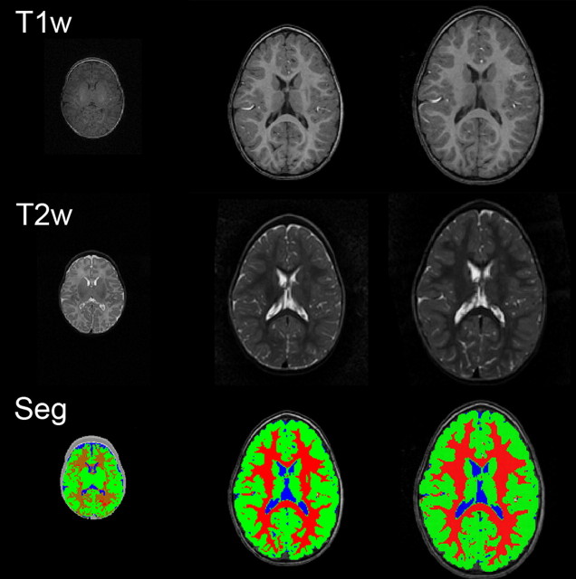
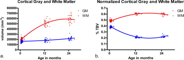
Brain development in the first 2 years after birth is extremely dynamic and likely plays an important role in neurodevelopmental disorders, including autism and schizophrenia. Knowledge regarding this period is currently quite limited. We studied structural brain development in healthy subjects from birth to 2. Ninety-eight children received structural MRI scans on a Siemens head-only 3T scanner with magnetization prepared rapid gradient echo T1-weighted, and turbo spin echo, dual-echo (proton density and T2 weighted) sequences: 84 children at 2-4 weeks, 35 at 1 year and 26 at 2 years of age. Tissue segmentation was accomplished using a novel automated approach. Lateral ventricle, caudate, and hippocampal volumes were also determined. Total brain volume increased 101% in the first year, with a 15% increase in the second. The majority of hemispheric growth was accounted for by gray matter, which increased 149% in the first year; hemispheric white matter volume increased by only 11%. Cerebellum volume increased 240% in the first year. Lateral ventricle volume increased 280% in the first year, with a small decrease in the second. The caudate increased 19% and the hippocampus 13% from age 1 to age 2. There was robust growth of the human brain in the first two years of life, driven mainly by gray matter growth. In contrast, white matter growth was much slower. Cerebellum volume also increased substantially in the first year of life. These results suggest the structural underpinnings of cognitive and motor development in early childhood, as well as the potential pathogenesis of neurodevelopmental disorders.
Figures

References
-
- Aljabar P, Bhatia KK, Murgasova M, Hajnal JV, Boardman JP, Srinivasan L, Rutherford MA, Dyet LE, Edwards AD, Rueckert D. Assessment of brain growth in early childhood using deformation-based morphometry. Neuroimage. 2008;39:348–358. - PubMed
-
- Allen NJ, Barres BA. Signaling between glia and neurons: focus on synaptic plasticity. Curr Opin Neurobiol. 2005;15:542–548. - PubMed
-
- Bailey A, Luthert P, Bolton P, Le Couteur A, Rutter M. Autism and megalencephaly. Lancet. 1993;341:1225–1226. - PubMed
-
- Bastian A, Thach WT. Structure and function of the cerebellum. In: Manto M, Pandolfo M, editors. The cerebellum and its disorders. Cambridge, UK: Cambridge UP; 2002. pp. 49–66.
-
- Benes FM, Turtle M, Khan Y, Farol P. Myelination of a key relay zone in the hippocampal-formation occurs in the human brain during childhood, adolescence, and adulthood. Arch Gen Psychiatry. 1994;51:477–484. - PubMed
Publication types
MeSH terms
Grants and funding
LinkOut - more resources
Full Text Sources
Other Literature Sources
Medical