. 2008 Nov 19;28(47):12328-40.
doi: 10.1523/JNEUROSCI.4000-08.2008.

Specific glial populations regulate hippocampal morphogenesis

Affiliations

Specific glial populations regulate hippocampal morphogenesis

Guy Barry et al. J Neurosci. .

Abstract

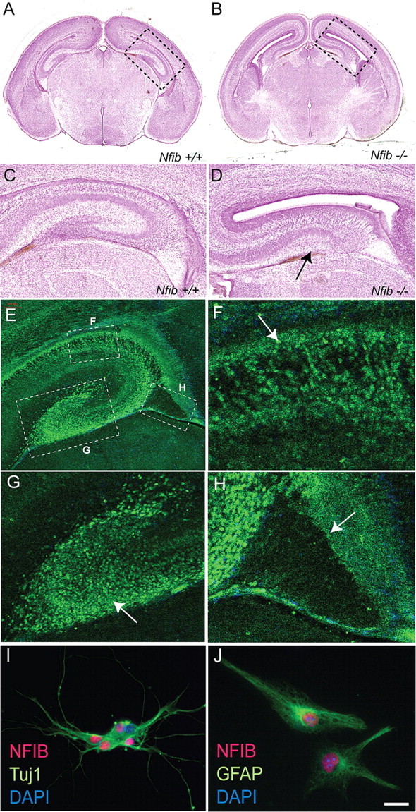
The hippocampus plays an integral role in spatial navigation, learning and memory, and is a major site for adult neurogenesis. Critical to these functions is the proper organization of the hippocampus during development. Radial glia are known to regulate hippocampal formation, but their precise function in this process is yet to be defined. We find that in Nuclear Factor I b (Nfib)-deficient mice, a subpopulation of glia from the ammonic neuroepithelium of the hippocampus fail to develop. This results in severe morphological defects, including a failure of the hippocampal fissure, and subsequently the dentate gyrus, to form. As in wild-type mice, immature nestin-positive glia, which encompass all types of radial glia, populate the hippocampus in Nfib-deficient mice at embryonic day 15. However, these fail to mature into GLAST- and GFAP-positive glia, and the supragranular glial bundle is absent. In contrast, the fimbrial glial bundle forms, but alone is insufficient for proper hippocampal morphogenesis. Dentate granule neurons are present in the mutant hippocampus but their migration is aberrant, likely resulting from the lack of the complete radial glial scaffold usually provided by both glial bundles. These data demonstrate a role for Nfib in hippocampal fissure and dentate gyrus formation, and that distinct glial bundles are critical for correct hippocampal morphogenesis.


Figures

Figure 1.
Figure 1.
NFIB is expressed in hippocampal neurons and glia. A–D, Hematoxylin staining of coronal sections of E18 wild-type (A, C) and Nfib-deficient (B, D) mouse brains. The DG is morphologically absent in the hippocampus of Nfib-deficient mice (D). The point at which the hippocampal fissure has failed to form in the mutant is indicated with an arrow in D. Panels C and D are higher magnifications of the boxed regions in A and B, respectively. E–H, Coronal section of an E18 wild-type brain (E), showing expression of NFIB protein in the VZ of the hippocampus (arrow in F) and in the DG (arrow in G). Only low levels of NFIB are expressed in the fimbria (arrow in H). I–J, In vitro cultures of wild-type hippocampal cells demonstrating that NFIB colocalizes with both the neuronal marker TuJ1 (I) and the glial marker GFAP (J). Scale bar: A, B, 500 μm; C, D, E, 80 μm; F–H, 25 μm; I, 60 μm; J, 15 μm.
Figure 2.
Figure 2.
Nestin-positive radial glia are present in Nfib-deficient mice. A, The stages of glial differentiation can be followed through the temporal expression of specific molecules including nestin (E11 onwards), GLAST (E13 onwards) and GFAP (E15 onwards). B–G, Expression of the radial progenitor marker nestin at E14 (B, C), E16 (D, E), and E18 (F, G). Nestin is expressed in wild-type (B, D, F) and Nfib-deficient mice (C, E, G), demonstrating that radial progenitors are specified in the absence of Nfib. Expression of nestin appears higher at E18 in the mutant (arrowhead in G). Arrows in B and C indicate the site of the future hippocampal fissure. Scale bar: B, C, 200 μm; D, E, 125 μm; F, G, 80 μm.
Figure 3.
Figure 3.
Aberrant expression of GLAST and tenascin-C in the hippocampus of Nfib-deficient mice. A–H, Expression of the glial markers GLAST (A–F) and tenascin-C (G, H) in coronal sections of wild-type (A, C, E, G) and Nfib-deficient (B, D, F, H) mice. GLAST expression is evident at E14 in the ammonic neuroepithelium of the wild-type hippocampus (A) but, strikingly, is absent from the ammonic neuroepithelium of Nfib null mutants (B), although it is evident in the fimbria (arrowheads in B, D, F). Arrows indicate GLAST-positive fibers converging at the future “anchor-point” of the hippocampal fissure. Asterisks indicate GLAST expression in the meninges. At E18, expression of GLAST in the mutant is present, but at a lower level than in the wild-type (compare E to F). Similarly, expression of tenascin-C in the mutant (H) is markedly lower than in the wild type (G) at E15. Scale bar: A, B, 200 μm; C, D, G, H, 180 μm; E, F, 80 μm.
Figure 4.
Figure 4.
The supragranular glial bundle fails to form in Nfib knock-out mice. A–H, Coronal sections of the hippocampus between E15 and E18 in wild-type (A, C, E, G) and Nfib-deficient (B, D, F, H) mice, showing expression of GFAP, a marker for mature radial glia. At E15, expression in the wild type (A) is evident in the fimbrial glioepithelium (arrowhead) and ammonic neuroepithelium (arrow), but is only seen in the fimbrial glioepithelium of the mutant (arrowhead in B). At E16, the disparity in GFAP expression in the ammonic neuroepithelium between the wild type (C) and knock-out (D) becomes more apparent. The boxed regions in C and D are shown at a higher magnification in E and F, respectively. In the wild type (E), both the fimbrial bundle (arrowhead) and the supragranular bundle (arrow) are evident, whereas only the fimbrial bundle is detected in the mutant (arrowhead in F). At E18, GFAP is expressed broadly in the wild-type hippocampus (G). Notably, a low level of GFAP expression is observed in the mutant ammonic neuroepithelium at E18 (arrow in H), indicating that GFAP expression may be delayed or compensated for by another mechanism. Scale bar: A, B, 180 μm; C, D, 125 μm; E, F, 40 μm; G, H, 80 μm.
Figure 5.
Figure 5.
Cultured hippocampal radial glia from Nfib-deficient mice display an altered morphology. Dissociated wild-type hippocampal cells were cultured for 24 h in vitro. A, Immunohistochemistry against the radial progenitor marker nestin demonstrated that under culture conditions all cells expressed this protein. Furthermore, 85% were also NFIB-positive (arrow indicates an NFIB-positive cell, arrowhead indicates an NFIB-negative cell). B, C, Hippocampal cells from wild-type (B) and mutant (C) mice were cultured for 24 h. Staining with phalloidin, a marker for filamentous actin, demonstrated that cells lacking Nfib exhibited an aberrant morphology. Analysis of both 24 and 48 h cultures showed that, although the length of the longest process was similar between wild-type (WT) and knock-out (KO) cells (D), cells from Nfib-deficient hippocampi had significantly more processes emanating from the cell body (E) and significantly more process branch points (F) in comparison with wild-type controls. *p < 0.0001, Student's t test. Error bars indicate SEM. G–J, Coronal paraffin sections of wild-type (G, I) and Nfib-deficient brains (H, J) at E17 showing expression of nestin. The boxed regions in G and H are shown at a higher magnification in I and J, respectively. The expression of nestin in knock-outs appears higher than that in controls, and, furthermore, whereas nestin fibers appear straight in the wild type (arrowhead in I), many fibers in the mutant appear wavy (arrow in J). Scale bars: (in C) A, 40 μm; B, C, 10 μm; (in J) G, H, 100 μm; I, J, 15 μm.
Figure 6.
Figure 6.
Aberrant proliferation in the hippocampus of Nfi-deficient mice at E18. Phosphohistone H3 (PH3) immunohistochemistry on wild-type (A) and Nfib-deficient mice (B) in the mouse hippocampus at E18. The number of PH3-positive cells in the ventricular zone (VZ) of the hippocampus was higher in Nfib-deficient mice compared with controls, whereas there were fewer PH3-positive cells in the dentate migratory stream (indicated by dashed lines) of the mutant. Scale bars: A, B, 80 μm.
Figure 7.
Figure 7.
Defects in the migration of dentate granule cells in Nfib-deficient mice. A–D, Coronal sections of wild-type (A, C) and Nfib-deficient mice (B, D) at E16 (A, B) and E18 (C, D) labeled with Prox1, a marker for dentate granule cells. Prox1-positive granule cells are born in the dentate neuroepithelium (arrowheads) of both wild-type (A) and Nfib-deficient (B) mice but fail to migrate into the presumptive DG (arrow in C) in mice lacking Nfib (D). Quantification of the number of Prox1-positive cells in 6 μm coronal paraffin sections showed no significant difference between E17 wild-type (E, 168.7 ± 12.91 cells) and Nfib-deficient (F, 162.3 ± 4.41 cells) hippocampal mouse sections. Statistical error indicates SEM. Scale bar: A, B, 125 μm; C, D, 80 μm.E,F 100 μm.
Figure 8.
Figure 8.
Glutamatergic neurons develop in the hippocampus of Nfi-deficient mice. A–F, Expression of Tbr1, a marker for glutamatergic neurons, in coronal sections of wild-type (A, C, E) and Nfib-deficient (B, D, F) hippocampi at E14 (A, B), E16 (C, D) and E18 (E, F). Tbr1-positive pyramidal neurons are generated and migrate within the hippocampus of Nfib-deficient mice. However, some notable differences between wild types and mutants are apparent. Lamination of the hippocampus is first observed at E16 in the wild type (C, arrows) but only at E18 in the mutant (F, arrows). Furthermore, the migration of Tbr1-positive neurons into the developing DG observed in the wild type (E) is aberrant in the mutant (F). G–J, Expression of the CA1-specfic marker SCIP (G, H) and the CA3-specific marker KA1 (I–J) visualized using in situ hybridization. The specification and differentiation of both CA1 and CA3 hippocampal subfields is apparent in Nfib-deficient mice. Scale bar: A, B, 200 μm; C, D, 125 μm; E–J, 80 μm.
Figure 9.
Figure 9.
Migration of interneurons and Cajal-Retzius cells occurs normally in Nfib-deficient mice. A–J, Expression of calbindin (A–D), calretinin (E–H) and reelin (I, J) in coronal sections of wild-type and Nfib-deficient mice. Calbindin-positive interneurons migrate normally into the hippocampus of mice lacking Nfib (B, D). At E16, calretinin expression is evident in the marginal zone of both wild-type (E) and mutant (F) hippocampi. However, the developing hippocampal fissure (arrow in E) is morphologically absent in the mutant. Comparison of wild-type (G) and mutant (H) sections at E18 demonstrates the absence of the DG (arrowhead in G) in mice lacking Nfib. Migration of reelin-positive Cajal-Retzius cells (I, J) into the hippocampus also occurs normally. However, Cajal-Retzius cells fail to migrate dorsally from the marginal zone into the DG by E18 in the mutant (J). Scale bar: A, B, E, F, 125 μm; C, D, G–J, 80 μm.
Figure 10.
Figure 10.
Summary of hippocampal defects in Nfib knock-out mice. A, B, Differentiating radial glial cells are depicted in the hippocampus of wild-type (A) or Nfib-deficient mice (B). At E18 in the wild-type hippocampus, the GFAP-expressing supragranular and fimbrial glial bundles have formed and the DG is developing its characteristic V-shape (A). Incomplete glial development occurs in Nfib-deficient mice, resulting in the formation of the fimbrial glial bundle but not the supragranular glial bundle, thus severely disrupting hippocampal development (B). These results demonstrate that the correct morphogenesis of the DG requires both GFAP-expressing glial bundles.

References

    1. Altman J, Bayer SA. Migration and distribution of two populations of hippocampal granule cell precursors during the perinatal and postnatal periods. J Comp Neurol. 1990a;301:365–381. - PubMed
    1. Altman J, Bayer SA. Mosaic organization of the hippocampal neuroepithelium and the multiple germinal sources of dentate granule cells. J Comp Neurol. 1990b;301:325–342. - PubMed
    1. Bettler B, Boulter J, Hermans-Borgmeyer I, O'Shea-Greenfield A, Deneris ES, Moll C, Borgmeyer U, Hollmann M, Heinemann S. Cloning of a novel glutamate receptor subunit, GluR5: expression in the nervous system during development. Neuron. 1990;5:583–595. - PubMed
    1. Bielle F, Griveau A, Narboux-Nême N, Vigneau S, Sigrist M, Arber S, Wassef M, Pierani A. Multiple origins of Cajal-Retzius cells at the borders of the developing pallium. Nat Neurosci. 2005;8:1002–1012. - PubMed
    1. Bulchand S, Grove EA, Porter FD, Tole S. LIM-homeodomain gene Lhx2 regulates the formation of the cortical hem. Mech Dev. 2001;100:165–175. - PubMed

Publication types

MeSH terms