. 2008 Nov 19;28(47):12427-32.
doi: 10.1523/JNEUROSCI.3573-08.2008.

The Semaphorin receptor PlexinA3 mediates neuronal apoptosis during dorsal root ganglia development

Affiliations

The Semaphorin receptor PlexinA3 mediates neuronal apoptosis during dorsal root ganglia development

Ayal Ben-Zvi et al. J Neurosci. .

Abstract

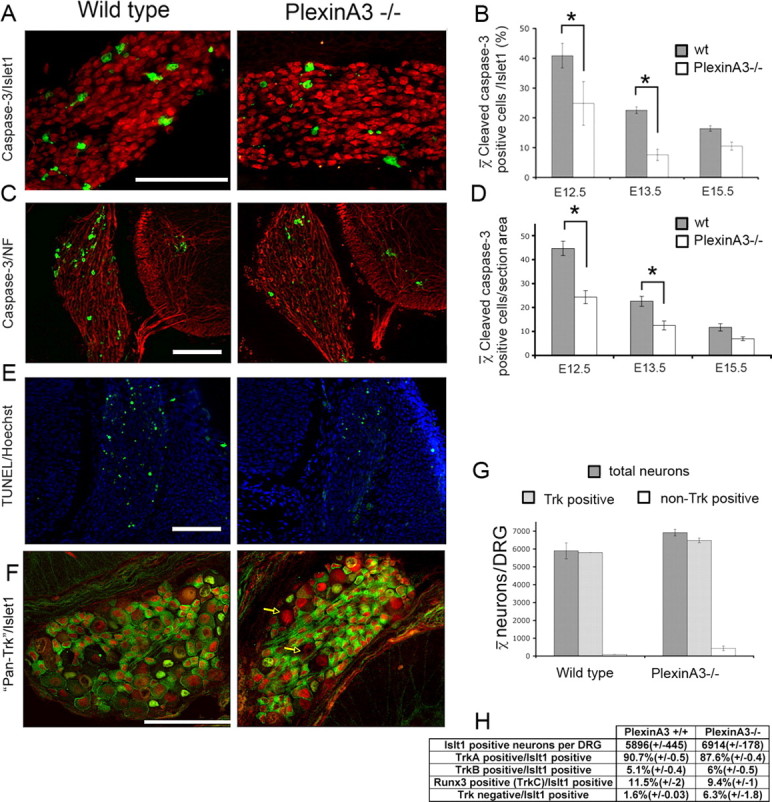
Extensive neuronal cell death during development is believed to be due to a limiting supply of neurotrophic factors. In vitro studies suggest that axon guidance molecules directly regulate neuronal survival, raising the possibility that they play a direct role in neuronal cell death in vivo. However, guidance errors may also influence survival indirectly due to loss of target-derived neurotrophic support. The role of guidance molecules in neuronal death in vivo has thus been difficult to decipher. Semaphorin3A, a repulsive guidance cue for sensory neurons, can induce sensory neuron death in vitro. Null mice studies of the Semaphorin3A coreceptors showed that guidance activity is mediated by PlexinA4, but PlexinA3 partially compensates in PlexinA4(-/-) mice. Here we demonstrate that both Plexins contribute to Sema3A-induced cell death in vitro, albeit in a different hierarchy. PlexinA3 is absolutely required, while PlexinA4 makes a smaller contribution to cell death. We found that PlexinA3(-/-) mice, which, unlike PlexinA4(-/-) mice, do not exhibit sensory axon patterning defects, show reduced neuronal apoptosis and an increased number of DRG neurons. Semaphorin3A involvement in neuronal death in vivo was demonstrated by a sensitization experiment using the proapoptotic effector Bax. Our results identify Plexins as mediators of Semaphorin-induced cell death in vitro, and provide the first evidence implicating Semaphorin/Plexin signaling in neuronal survival independent of its role in axon guidance. The results also support the idea that naturally occurring neuronal cell death reflects not only competition for target-derived trophic factors, but also the action of proapoptotic signaling via a Semaphorin/Plexin pathway.


Figures

Figure 1.
Figure 1.
PlexinA3 is the key receptor subunit responsible for Sema3A-induced cell death. Survival effects of Sema3A on NGF-dependent neuronal cultures prepared from L1−/−, PlexinA3−/−, or PlexinA4−/− mice. Neurons from wild-type littermates were prepared for each mutant line. In each experiment, neurons were grown for 16 h and then re-fed (t = 0) with fresh medium in the presence or absence of 100 pm Sema3A. The same fields were counted at t = 0, and t = 24 h. A, Representative cultures of wild-type (top) or PlexinA3−/− (bottom) neurons are shown at time = 0 and 24 h of the same field. Note the changes in the neurite network in PlexinA3−/− neuronal cultures because of additional Sema3A functions that are not related to its cell death activity and are not blocked in the absence of this receptor component. Arrows indicate examples of surviving neurons and arrowheads marks examples of neurons at t = 0 h that are no longer present at t = 24 h. B, Sema3A-induced reduction in cell survival of wild-type neurons (for each experiment) was defined as a 100% killing-effect. The killing-effect (%) in mutant embryos normalized to wild-type neurons is presented for each mouse line (C) NT-3- and BDNF-dependent neuronal cultures were prepared from PlexinA3−/− and wild-type littermates, and the killing-effect (%) of Sema3A was calculated as in B. D, Survival of neurons (PlexinA3−/− and wild-type littermates) following neurotrophin withdrawal (NGF- and NT-3-dependent neuronal cultures) was tested. Neurons were grown for 16 h before the medium was washed and replaced (t = 0) with fresh medium with or without neurotrophins. The same fields were counted at each time point. The percentage of surviving neurons in neurotrophin withdrawal experiments is normalized at each time point to percentage of neuronal survival in cultures with neurotrophins. Differences between wild-type and PlexinA3−/− neurons are significant (*p < 0.014, χ2 test). In all experiments, results represents mean ± SEM of three independent experiments. Scale bar, 100 μm.
Figure 2.
Figure 2.
Loss of PlexinA3 results in a reduction in DRG cell death in vivo. E12.5, E13.5 and E15.5 embryos (PlexinA3−/− and wild-type littermates) were fixed and sectioned (A, B). A, Sections from E12.5, E13.5 and E15.5 embryos (PlexinA3−/− and wild-type littermates) were stained with anti-cleaved caspase-3 (green) and anti-Islet-1 (red). Representative microscopic fields from E13.5 wild-type and PlexinA3−/− embryos are shown. B, Quantification of the average cleaved caspase-3-positive cells/Islet-1-positive cells (%) is also shown. This difference is significant both in E12.5 and E13.5 (p < 10−5). C, Sections taken from E13.5 PlexinA3−/− and wild-type littermates stained with anti-cleaved caspase-3 (green) and antineurofilament (red) are shown, together with their quantification (D). The average number of cleaved caspase-3-positive cells/section area in PlexinA3 mutants is significantly lower (E12.5 p = 0.016, E13.5 p = 0.05) than in wild-type littermates. E, TUNEL assay of E12.5 lumbar sections from PlexinA3−/− and PlexinA3+/+ littermates. TUNEL-positive cells are labeled in green. Nuclei are labeled with Hoechst (blue). F, Example of sections taken from E15.5 PlexinA3−/− and wild-type littermates stained with a mixture of anti-TrkA, anti-TrkB, and anti-RUNX3 (green) in conjunction with anti-Islet-1 (red). Arrows indicate Islet-1-positive/Trk-negative neurons. G, H, Total number of neurons and percentage of each subpopulation (TrkA, TrkB or RUNX3) are shown. (For reasons of clarity, the Trk-negative and Trk-positive percentages in G were translated into numbers using the total neuronal number counts.) Results represent mean ± SEM. Scale bars, 100 μm.
Figure 3.
Figure 3.
Genetic sensitization experiment using BAX+/− reveals the involvement of Sema3A-induced death in the developing DRG. E12.5 embryos (Sema3A−/−;BAX+/−, Sema3A−/−;BAX+/+, Sema3A+/+BAX+/−, and Sema3A+/+;BAX+/+ littermates) were fixed and sectioned. Quantification of cleaved caspase-3-positive cells is shown. The number of cleaved caspase-3-positive cells in Sema3A−/− that are also heterozygous for BAX gene is significantly lower that BAX+/− only embryos (p = 0.034, Mann–Whitney U test).

References

    1. Avivi C, Goldstein RS. Differential expression of Islet-1 in neural crest-derived ganglia: Islet-1 + dorsal root ganglion cells are post-mitotic and Islet-1 + sympathetic ganglion cells are still cycling. Brain Res Dev Brain Res. 1999;115:89–92. - PubMed
    1. Bagri A, Cheng HJ, Yaron A, Pleasure SJ, Tessier-Lavigne M. Stereotyped pruning of long hippocampal axon branches triggered by retraction inducers of the semaphorin family. Cell. 2003;113:285–299. - PubMed
    1. Behar O, Golden JA, Mashimo H, Schoen FJ, Fishman MC. Semaphorin III is needed for normal patterning and growth of nerves, bones and heart. Nature. 1996;383:525–528. - PubMed
    1. Ben-Zvi A, Yagil Z, Hagalili Y, Klein H, Lerman O, Behar O. Semaphorin 3A and neurotrophins: a balance between apoptosis and survival signaling in embryonic DRG neurons. J Neurochem. 2006;96:585–597. - PubMed
    1. Burek MJ, Oppenheim RW. Programmed cell death in the developing nervous system. Brain Pathol. 1996;6:427–446. - PubMed

Publication types

MeSH terms