. 2008 Dec 2;105(48):18924-9.
doi: 10.1073/pnas.0810230105. Epub 2008 Nov 19.

Role of nitric oxide signaling components in differentiation of embryonic stem cells into myocardial cells

Affiliations

Role of nitric oxide signaling components in differentiation of embryonic stem cells into myocardial cells

Kalpana Mujoo et al. Proc Natl Acad Sci U S A. .

Abstract

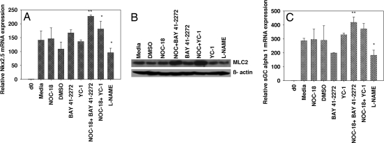
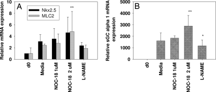
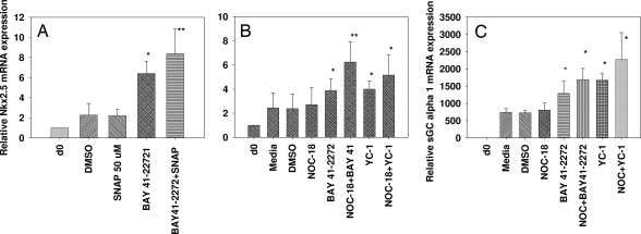
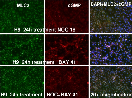
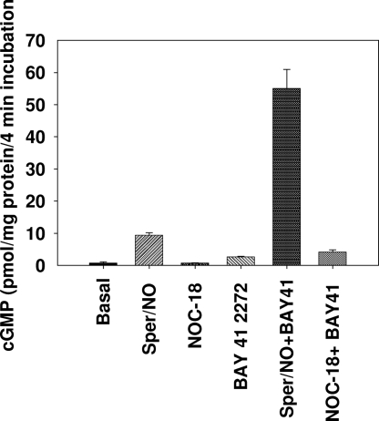
Nitric oxide (NO) is involved in number of physiological and pathological events. Our previous studies demonstrated a differential expression of NO signaling components in mouse and human ES cells. Here, we demonstrate the effect of NO donors and soluble guanylyl cyclase (sGC) activators in differentiation of ES cells into myocardial cells. Our results with mouse and human ES cells demonstrate an increase in Nkx2.5 and myosin light chain (MLC2) mRNA expression on exposure of cells to NO donors and a decrease in mRNA expression of both cardiac-specific genes with nonspecific NOS inhibitor and a concomitant increase and decrease in the mRNA levels of sGC alpha(1) subunit. Although sGC activators alone exhibited an increase in mRNA expression of cardiac genes (MLC2 and Nkx2.5), robust inductions of mRNA and protein expression of marker genes were observed when NO donors and sGC activators were combined. Measurement of NO metabolites revealed an increase in the nitrite levels in the conditioned media and cell lysates on exposure of cells to the different concentrations of NO donors. cGMP analysis in undifferentiated stem cells revealed a lack of stimulation with NO donors. Differentiated cells however, acquired the ability to be stimulated by NO donors. Although, 3-(4-amino-5-cyclopropylpyrimidin-2-yl)-1-(2-fluorobenzyl)-1H-pyrazolo [3,4-b]pyridine (BAY 41-2272) alone was able to stimulate cGMP accumulation, the combination of NO donors and BAY 41-2272 stimulated cGMP levels more than either of the agents separately. These studies demonstrate that cGMP-mediated NO signaling plays an important role in the differentiation of ES cells into myocardial cells.


Conflict of interest statement

The authors declare no conflict of interest.

Figures

Fig. 1.
Fig. 1.
Effect of an NO donor, sGC activators, and a NOS inhibitor on EZ1 (murine ES) cells. The EZ1 cells were differentiated and exposed to NOC-18 (1 μM), BAY 41-2272, YC-1 (3 μM), l- NAME (1 mM), and a combination of NOC + BAY 41-2272. or NOC + YC-1 on days 0, 5, and 7 and harvested on day 10. (A and C) mRNA levels for the indicated genes were analyzed by real-time PCR and normalized by using the housekeeping gene GAPDH and presented as fold expression compared with day 0. Day 0 represents undifferentiated cell culture usually collected before subjecting cells to differentiation. The data were analyzed by using the 2−ΔΔCt method. Error bars indicate ± SEM. Significance using paired Student's t test is indicated: *, P < 0.05; **, P < 0.01. n = 3. (B) The protein from EZ1 cells untreated or treated with NOC-18 (1 μM), BAY 41-2272, YC-1 (3 μM), l- NAME (1 mM), or the combination exposed on days 0, 5, and 7 and harvested on day 10 was detected with specific antibodies against MLC2 and β-actin by using Western blot–ECL analysis.
Fig. 2.
Fig. 2.
Effect of NOC-18 and l-NAME in H-9 cells. Human ES cells (H-9) were subjected to differentiation by using the EB method, and the differentiated cells were exposed to an NO donor, NOC-18 (1–2 μM), and a nonspecific NOS inhibitor, l-NAME (2 mM), on day 17 for 24 h. mRNA expression for the indicated genes (MLC2, NkX2.5, sGC α1, and GAPDH) was analyzed by real-time PCR. All of the samples were normalized by using housekeeping gene GAPDH, and data were analyzed by the 2−ΔΔCt method. Error bars (±SEM) indicate significance using paired Student's t test is indicated. *, P < 0.05; **, P < 0.01; n = 4.
Fig. 3.
Fig. 3.
Effect of BAY 41-2272 on Nkx2.5 mRNA on H-9 cells. (A and B) Partially differentiated H-9 cells were exposed to a single treatment on day 13 (A) or multiple treatments (starting on day 7, total number of exposures 4 (B). The cells were harvested on day 14, and mRNA expression of Nkx2.5 was determined by real-time PCR. All of the samples were normalized with the housekeeping gene GAPDH. *, P < 0.05; **, P < 0.01; n = 3–6. (C) The protein from differentiated H-9 cells either untreated or treated with BAY 41-2272 (1–3 μM) or ascorbic acid (30–90 μM) on days 7, 9, 11, and 13 and harvested on day 14 was extracted and detected with specific antibodies against MLC2 and β-actin by using Western blot–ECL analysis.
Fig. 4.
Fig. 4.
Effect of NO donors and sGC activators in H-9 cells. Partially differentiated cells were exposed to a single treatment of the NO donor SNAP (50 μM), BAY 41-2272 (5 μM), and the combination of SNAP+BAY41 (A) or multiple treatments of NOC-18 (1 μM), BAY 41-2272 (3 μM), or the combination of NOC-18+BAY41, YC-1 (3 μM) or the combination of NOC-18 + YC-1 (B and C). The samples were analyzed for Nkx2.5 (A and B) and sGC α1 transcript levels (C) by quantitative RT-PCR. Error bars indicate ± SEM. Significance using paired Student's t test is shown: *, P < 0.05; **, P < 0.01. n = 5–7.
Fig. 5.
Fig. 5.
Immunofluorescence detection of MLC2 and cGMP in H-9 cells exposed to NO donor and sGC activator. Differentiated cells were exposed to NOC-18 (1 μM; Top), BAY 41-2272 (3 μM; Middle), or the combination of the two (C) for 24 h at 37 °C. The cells were fixed with paraformaldehyde and incubated with antibodies to MLC2 and cGMP that was followed by detection with fluorescent-conjugated secondary anti-mouse or rabbit antibodies. (Magnification: 20×.)
Fig. 6.
Fig. 6.
cGMP accumulation in undifferentiated and differentiated H-9 cells. H-9 cells were harvested with 1 mM EDTA in DPBS and incubated for 10 min at 37 °C in DPBS containing 1 mM IBMX with 0.1% DMSO or 5 μM BAY 41-2272 (2–3 × 10⁷ cells per mL) followed by the addition of NO donors. Cells were incubated for 4 min and then 50 μL of ice-cold 1 M perchloric acid was added and cGMP was determined by ELISA. Data are expressed in pmol cGMP per mg of protein.

References

    1. Thomson JA, et al. Embryonic stem cell lines derived from human blastocysts. Science. 1997;282:1145–1147. - PubMed
    1. Reubinoff BE, Pera MF, Fong CY, Trounson A, Bongso A. Embryonic stem cell lines from human blastocysts: Somatic differentiation in vitro. Nat Biotechnol. 2000;18:399–404. - PubMed
    1. Hescheler J, et al. Embryonic stem cells: A model to study structural and functional properties in cardiomyogenesis. Cardiovasc Res. 1997;36:149–162. - PubMed
    1. Min JY, et al. Transplantation of embryonic stem cells improves cardiac function in postinfarcted rats. J Appl Physiol. 2002;92:288–296. - PubMed
    1. Min JY, et al. Long-term improvement of cardiac function in rats after infarction by transplantation of embryonic stem cells. J Thorac Cardiovasc Surg. 2003;125:361–369. - PubMed

Publication types

MeSH terms

LinkOut - more resources