. 2008 Nov 25;105(47):18531-6.
doi: 10.1073/pnas.0808010105. Epub 2008 Nov 19.

A bifunctional kinase-phosphatase in bacterial chemotaxis

Affiliations

A bifunctional kinase-phosphatase in bacterial chemotaxis

Steven L Porter et al. Proc Natl Acad Sci U S A. .

Abstract

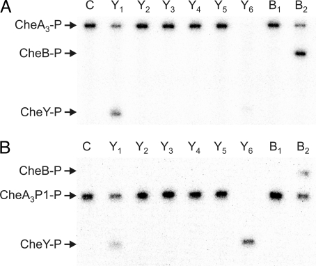
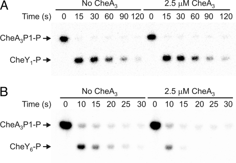
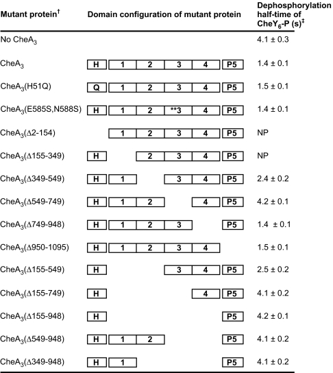
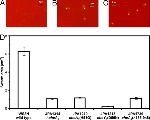
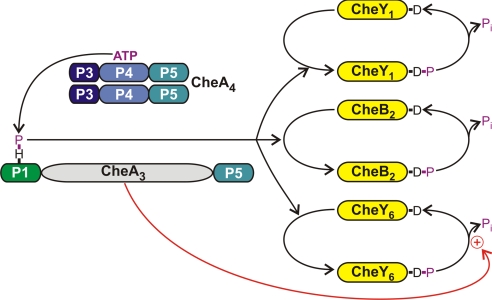
Phosphorylation-based signaling pathways employ dephosphorylation mechanisms for signal termination. Histidine to aspartate phosphosignaling in the two-component system that controls bacterial chemotaxis has been studied extensively. Rhodobacter sphaeroides has a complex chemosensory pathway with multiple homologues of the Escherichia coli chemosensory proteins, although it lacks homologues of known signal-terminating CheY-P phosphatases, such as CheZ, CheC, FliY or CheX. Here, we demonstrate that an unusual CheA homologue, CheA(3), is not only a phosphodonor for the principal CheY protein, CheY(6), but is also is a specific phosphatase for CheY(6)-P. This phosphatase activity accelerates CheY(6)-P dephosphorylation to a rate that is comparable with the measured stimulus response time of approximately 1 s. CheA(3) possesses only two of the five domains found in classical CheAs, the Hpt (P1) and regulatory (P5) domains, which are joined by a 794-amino acid sequence that is required for phosphatase activity. The P1 domain of CheA(3) is phosphorylated by CheA(4), and it subsequently acts as a phosphodonor for the response regulators. A CheA(3) mutant protein without the 794-amino acid region lacked phosphatase activity, retained phosphotransfer function, but did not support chemotaxis, suggesting that the phosphatase activity may be required for chemotaxis. Using a nested deletion approach, we showed that a 200-amino acid segment of CheA(3) is required for phosphatase activity. The phosphatase activity of previously identified nonhybrid histidine protein kinases depends on the dimerization and histidine phosphorylation (DHp) domains. However, CheA(3) lacks a DHp domain, suggesting that its phosphatase mechanism is different from that of other histidine protein kinases.


Conflict of interest statement

The authors declare no conflict of interest.

Figures

Fig. 1.
Fig. 1.
Phosphorimages of SDS/PAGE gels showing phosphotransfer from (A) CheA3 and (B) CheA3P1 (the isolated P1 domain of CheA3) to the R. sphaeroides chemotaxis response regulators. CheA3 (4 μM) and CheA4 (10 μM) were preincubated together with 0.5 mM [γ-32P] ATP for 30 min. RRs (5 μM) were then added. Ten-microliter reaction samples were removed after 30 s and quenched in 10 μl of 3× SDS/EDTA loading dye. The samples were analyzed by SDS/PAGE and detected by phosphorimaging. Lane C shows a control reaction in which an equal volume of buffer was added instead of the RRs. The remaining lanes are labeled according to which RR was used; for example, CheY1 was used in the lane labeled Y1. Phosphotransfer is indicated by the appearance of phosphorylated RR and a reduction in the amount of (A) CheA3-P or (B) CheA3P1-P.
Fig. 2.
Fig. 2.
Phosphorimages of SDS/PAGE gels showing the response regulator dephosphorylation time courses. (A) 400 μM CheY1 was added to 2 μM CheA3P1-P in the absence (left half of gel) and presence of 2.5 μM CheA3 (right half of gel). (B) 400 μM CheY6 was added to 30 μM CheA3P1-P in the absence (left half of gel) and presence of 2.5 μM CheA3 (right half of gel). 10-μl reaction samples were taken at the time points indicated and quenched in 20 μl of 1.5× SDS/EDTA loading dye. The quenched samples were analyzed by SDS/PAGE and detected by phosphorimaging. ATP was not present in any of the reactions, so after the phosphotransfer reactions, which were completed before the first-time point, the only reaction occurring was RR-P dephosphorylation. As has been observed for E. coli CheA, a small fraction of CheA3P1-P (< 4%) failed to transfer phosphoryl groups to the RRs (42). Phosphatase activity is indicated by a reduction in CheY-P levels in the presence of 2.5 μM CheA3 when compared with those in the absence of CheA3 (seen in B but not in A).
Fig. 3.
Fig. 3.
The effect of CheA3 mutant proteins on the dephosphorylation rate of CheY6-P. Two and one-half μM of each CheA3 mutant protein was used in the phosphatase assays. The molar ratio of CheY6 to CheA3 mutant protein was 160:1. Each experiment was performed six times and mean values ± standard error are shown (values are rounded to two significant figures). NP, could not be overexpressed and purified.
Fig. 4.
Fig. 4.
The 794-amino acid region between the P1 and P5 domains of CheA3 is not required for CheA3 localization but is required for chemotaxis. (A) YFP fluorescence image of wild-type cells (strain WS8N). (B) YFP fluorescence image of JPA1425 (yfp-cheA3). (C) YFP fluorescence image of JPA1741 [yfp-cheA3(Δ155–948)]. (D) Swarm plate chemotaxis assay comparing the chemotactic ability of JPA1739 [cheA3(Δ155–948)] with wild-type (WS8N), nonchemotactic (JPA1314 and JPA1210) and nonmotile (JPA1213) strains. The swarm plates contained 100 μM propionate and were incubated for 48 h under aerobic conditions. Error bars show the standard error of the mean obtained from nine independent experiments.
Fig. 5.
Fig. 5.
The phosphorylation reactions involving CheA3. The domain structures of CheA3 and CheA4 are shown. The P1 domain of CheA3 is phosphorylated by a CheA4 dimer. CheA3-P then acts as a phosphodonor for either CheY1, CheY6, or CheB2. These RRs all autodephosphorylate. However, CheA3 acts as a phosphatase on CheY6-P (red arrow) and can accelerate the rate of dephosphorylation by at least a factor of 3 over the rate of autodephosphorylation.

References

    1. Stock AM, Robinson VL, Goudreau PN. Two-component signal transduction. Annu Rev Biochem. 2000;69:183–215. - PubMed
    1. Ulrich LE, Zhulin IB. MiST: A microbial signal transduction database. Nucl Acids Res. 2007;35:D386–D390. - PMC - PubMed
    1. Galperin MY. Structural classification of bacterial response regulators: Diversity of output domains and domain combinations. J Bacteriol. 2006;188:4169–4182. - PMC - PubMed
    1. Perego M, et al. Multiple protein-aspartate phosphatases provide a mechanism for the integration of diverse signals in the control of development in B. subtilis. Cell. 1994;79:1047–1055. - PubMed
    1. Hess JF, Oosawa K, Kaplan N, Simon MI. Phosphorylation of three proteins in the signalling pathway of bacterial chemotaxis. Cell. 1988;53:79–87. - PubMed

Publication types

Substances