

Frequent in-frame somatic deletions activate gp130 in inflammatory hepatocellular tumours
- PMID: 19020503
- PMCID: PMC2695248
- DOI: 10.1038/nature07475
Frequent in-frame somatic deletions activate gp130 in inflammatory hepatocellular tumours
Abstract
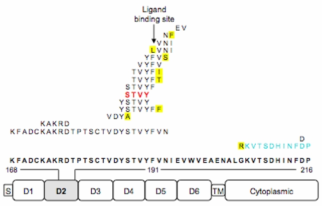
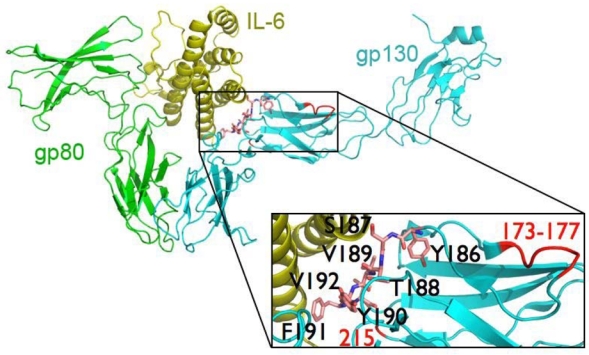
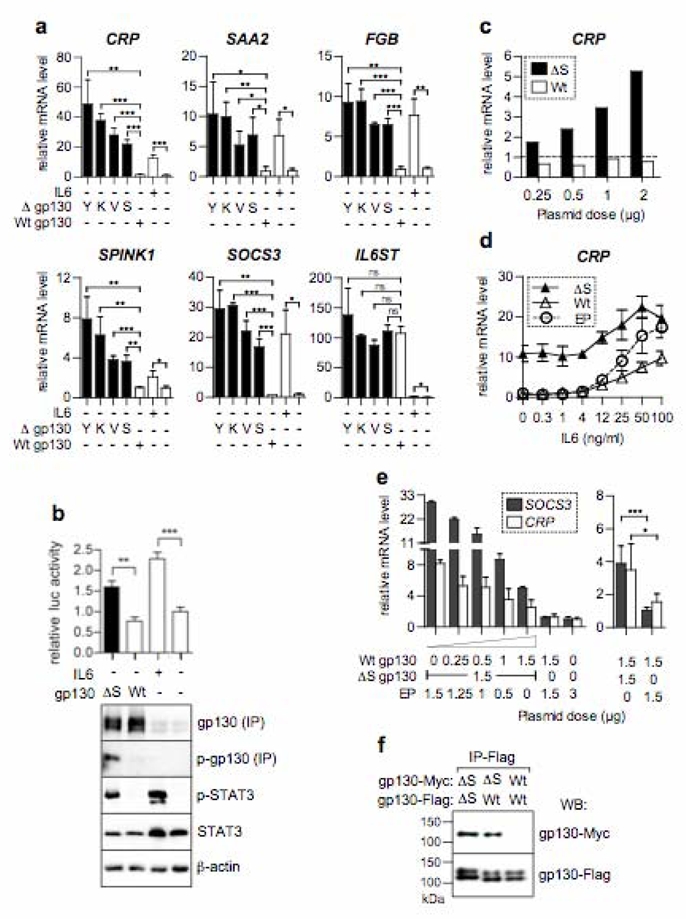
Inflammatory hepatocellular adenomas are benign liver tumours defined by the presence of inflammatory infiltrates and by the increased expression of inflammatory proteins in tumour hepatocytes. Here we show a marked activation of the interleukin (IL)-6 signalling pathway in this tumour type; sequencing candidate genes pinpointed this response to somatic gain-of-function mutations in the IL6ST gene, which encodes the signalling co-receptor gp130. Indeed, 60% of inflammatory hepatocellular adenomas harbour small in-frame deletions that target the binding site of gp130 for IL-6, and expression of four different gp130 mutants in hepatocellular cells activates signal transducer and activator of transcription 3 (STAT3) in the absence of ligand. Furthermore, analysis of hepatocellular carcinomas revealed that rare gp130 alterations are always accompanied by beta-catenin-activating mutations, suggesting a cooperative effect of these signalling pathways in the malignant conversion of hepatocytes. The recurrent gain-of-function gp130 mutations in these human hepatocellular adenomas fully explains activation of the acute inflammatory phase observed in tumourous hepatocytes, and suggests that similar alterations may occur in other inflammatory epithelial tumours with STAT3 activation.
Figures

Comment in
-
Frequent in-frame somatic deletions activate gp130 in inflammatory hepatocellular tumors.Hepatology. 2009 Apr;49(4):1387-9. doi: 10.1002/hep.22902. Hepatology. 2009. PMID: 19330860 No abstract available.
-
IL-6/gp130/STAT3 signaling axis in cancer and the presence of in-frame gp130 somatic deletions in inflammatory hepatocellular tumors.Future Oncol. 2009 Apr;5(3):305-8. doi: 10.2217/fon.09.3. Future Oncol. 2009. PMID: 19374537
-
A new link between cancer and inflammation?J Hepatol. 2009 Jul;51(1):230-2. doi: 10.1016/j.jhep.2009.03.004. Epub 2009 Mar 27. J Hepatol. 2009. PMID: 19446914 No abstract available.
Similar articles
-
Somatic mutations activating STAT3 in human inflammatory hepatocellular adenomas.J Exp Med. 2011 Jul 4;208(7):1359-66. doi: 10.1084/jem.20110283. Epub 2011 Jun 20. J Exp Med. 2011. PMID: 21690253 Free PMC article.
-
Inflammatory hepatocellular adenomas developed in the setting of chronic liver disease and cirrhosis.Mod Pathol. 2016 Jan;29(1):43-50. doi: 10.1038/modpathol.2015.119. Epub 2015 Oct 30. Mod Pathol. 2016. PMID: 26516697
-
Constitutively active mutant gp130 receptor protein from inflammatory hepatocellular adenoma is inhibited by an anti-gp130 antibody that specifically neutralizes interleukin 11 signaling.J Biol Chem. 2012 Apr 20;287(17):13743-51. doi: 10.1074/jbc.M111.349167. J Biol Chem. 2012. PMID: 22523320 Free PMC article.
-
Mutations leading to constitutive active gp130/JAK1/STAT3 pathway.Cytokine Growth Factor Rev. 2015 Oct;26(5):499-506. doi: 10.1016/j.cytogfr.2015.07.010. Epub 2015 Jul 3. Cytokine Growth Factor Rev. 2015. PMID: 26188635 Review.
-
The Human GP130 Cytokine Receptor and Its Expression-an Atlas and Functional Taxonomy of Genetic Variants.J Clin Immunol. 2023 Dec 22;44(1):30. doi: 10.1007/s10875-023-01603-7. J Clin Immunol. 2023. PMID: 38133879 Free PMC article. Review.
Cited by
-
The SphKs/S1P/S1PR1 axis in immunity and cancer: more ore to be mined.World J Surg Oncol. 2016 Apr 29;14:131. doi: 10.1186/s12957-016-0884-7. World J Surg Oncol. 2016. PMID: 27129720 Free PMC article. Review.
-
Oncogenic β-catenin triggers an inflammatory response that determines the aggressiveness of hepatocellular carcinoma in mice.J Clin Invest. 2012 Feb;122(2):586-99. doi: 10.1172/JCI43937. Epub 2012 Jan 17. J Clin Invest. 2012. PMID: 22251704 Free PMC article.
-
Expression of Carboxypeptidase X M14 Family Member 2 Accelerates the Progression of Hepatocellular Carcinoma via Regulation of the gp130/JAK2/Stat1 Pathway.Cancer Manag Res. 2020 Mar 31;12:2353-2364. doi: 10.2147/CMAR.S228984. eCollection 2020. Cancer Manag Res. 2020. PMID: 32280274 Free PMC article.
-
Synthetic Deletion of the Interleukin 23 Receptor (IL-23R) Stalk Region Led to Autonomous IL-23R Homodimerization and Activation.Mol Cell Biol. 2017 Aug 11;37(17):e00014-17. doi: 10.1128/MCB.00014-17. Print 2017 Sep 1. Mol Cell Biol. 2017. PMID: 28630278 Free PMC article.
-
LIF-independent JAK signalling to chromatin in embryonic stem cells uncovered from an adult stem cell disease.Nat Cell Biol. 2011 Jan;13(1):13-21. doi: 10.1038/ncb2135. Epub 2010 Dec 12. Nat Cell Biol. 2011. PMID: 21151131 Free PMC article.
References
-
- Bioulac-Sage P, et al. Hepatocellular adenoma subtype classification using molecular markers and immunohistochemistry. Hepatology. 2007;46:740–8. - PubMed
-
- Zucman-Rossi J, et al. Genotype-phenotype correlation in hepatocellular adenoma: new classification and relationship with HCC. Hepatology. 2006;43:515–24. - PubMed
-
- Grivennikov S, Karin M. Autocrine IL-6 signaling: a key event in tumourigenesis? Cancer Cell. 2008;13:7–9. - PubMed
-
- Akira S, et al. Molecular cloning of APRF, a novel IFN-stimulated gene factor 3 p91-related transcription factor involved in the gp130-mediated signaling pathway. Cell. 1994;77:63–71. - PubMed
Publication types
MeSH terms
Substances
Associated data
- Actions
Grants and funding
LinkOut - more resources
Full Text Sources
Other Literature Sources
Medical
Molecular Biology Databases
Miscellaneous
