

doi: 10.1007/978-1-59745-170-3_4.
Cell-free assays for HIV-1 uncoating
Affiliations
- PMID: 19020817
- PMCID: PMC3842014
- DOI: 10.1007/978-1-59745-170-3_4
Item in Clipboard
Cell-free assays for HIV-1 uncoating
Methods Mol Biol.
2009.
Abstract
Uncoating is an essential step in the retrovirus life cycle about which little is known. Uncoating is defined as the specific dissociation of the capsid shell from the viral core in the host cell cytoplasm. In this chapter, biochemical assays for studying HIV-1 uncoating in vitro are described. These techniques have proven useful for characterizing HIV-1 mutants that exhibit defects in the uncoating step of infection.
Figures

Schematic of HIV-1 uncoating. During incubation at 37°C, purified HIV-1 cores spontaneously release the CA and RT proteins into a soluble form. The extent of uncoating is determined by p24 ELISA after separating free from core-associated CA by ultracentifugation.
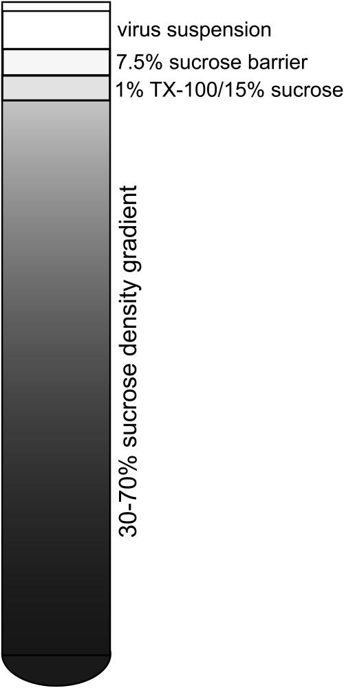
Construction of density gradients for isolation of HIV-1 cores. A 30-70% sucrose density gradient is prepared and successively overlaid with a layer of detergent, a barrier layer to prevent premature mixing of virus and detergent, and the concentrated virus suspension. Upon ultracentrifugation, virions pass through the detergent layer, releasing HIV-1 cores that then sediment to their equilibrium density.

Distribution of CA in the density gradient after ultracentifugation. Fractions from a gradient were collected from top to bottom and analyzed for p24 by ELISA and density by refractometry. In this experiment, a quantity of HIV-1 corresponding to approximately 250 μg of p24 was applied to the gradient.
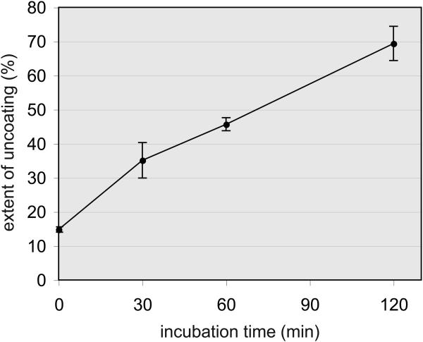
Uncoating of HIV-1 cores occurs spontaneously during incubation at 37°C. Samples of purified HIV-1 cores were diluted in buffer and incubated for the indicated times. Reactions were chilled and the cores were pelleted by ultracentrifugation. p24 concentrations in the pellets and supernatants were determined by ELISA, and the extent of uncoating was calculated. Error bars correspond to standard deviations from the mean values from triplicate reactions.
References
-
- Aiken C. Viral and cellular factors that regulate HIV-1 uncoating. Curr Opin HIV AIDS. 2006;1:194–199. - PubMed
-
- Wacharapornin P, Lauhakirti D, Auewarakul P. The effect of capsid mutations on HIV-1 uncoating. Virology. 2007;358:48–54. - PubMed
-
- Auewarakul P, Wacharapornin P, Srichatrapimuk S, Chutipongtanate S, Puthavathana P. Uncoating of HIV-1 requires cellular activation. Virology. 2005;337:93–101. - PubMed
Publication types
MeSH terms
Grants and funding
LinkOut - more resources
Full Text Sources
