. 2009 Sep;5(3):277-87.
doi: 10.1007/s11302-008-9128-9. Epub 2008 Nov 20.

Mouse Leydig cells express multiple P2X receptor subunits

Affiliations

Mouse Leydig cells express multiple P2X receptor subunits

Ligia Subitoni Antonio et al. Purinergic Signal. 2009 Sep.

Abstract

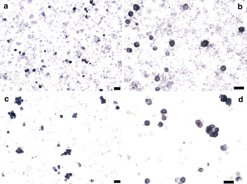
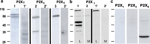
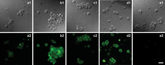
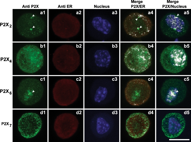
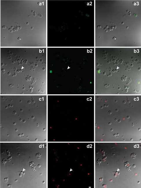
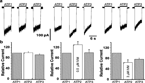
ATP acts on cellular membranes by interacting with P2X (ionotropic) and P2Y (metabotropic) receptors. Seven homomeric P2X receptors (P2X(1)-P2X(7)) and seven heteromeric receptors (P2X(1/2), P2X(1/4), P2X(1/5), P2X(2/3), P2X(2/6), P2X(4/6), P2X(4/7)) have been described. ATP treatment of Leydig cells leads to an increase in [Ca(2+)](i) and testosterone secretion, supporting the hypothesis that Ca(2+) signaling through purinergic receptors contributes to the process of testosterone secretion in these cells. Mouse Leydig cells have P2X receptors with a pharmacological and biophysical profile resembling P2X(2). In this work, we describe the presence of several P2X receptor subunits in mouse Leydig cells. Western blot experiments showed the presence of P2X(2), P2X(4), P2X(6), and P2X(7) subunits. These results were confirmed by immunofluorescence. Functional results support the hypothesis that heteromeric receptors are present in these cells since 0.5 muM ivermectin induced an increase (131.2 +/- 5.9%) and 3 muM ivermectin a decrease (64.2 +/- 4.8%) in the whole-cell currents evoked by ATP. These results indicate the presence of functional P2X(4) subunits. P2X(7) receptors were also present, but they were non-functional under the present conditions because dye uptake experiments with Lucifer yellow and ethidium bromide were negative. We conclude that a heteromeric channel, possibly P2X(2/4/6), is present in Leydig cells, but with an electrophysiological and pharmacological phenotype characteristic of the P2X(2) subunit.


Figures

Fig. 1
Fig. 1
Cytochemical detection of 3β-HSD on cell suspensions before and after hyposmotic shock. a and b The cell suspension was obtained by mechanical dispersion only and stained for 3β-HSD. c and d Mechanically isolated cells were also subjected to a hyposmotic shock and then stained for 3β-HSD. Leydig cells are marked in intense blue. Scale bar 20 μm
Fig. 2
Fig. 2
Western Blot analysis of purinergic receptors P2X1–7. a Western blot analysis of lysates of mouse Leydig cells performed with specific antibodies against rat P2X2 (lane 1), P2X4 (lane 2), and P2X6 (lane 3) receptors. Positively marked bands are seen at ∼66 kDa corresponding to these subunits. These same bands are absent in the experiments where the antibodies were pre-incubated with their respective cognate peptides (lane 1′, lane 2′, and lane 3′ for P2X2, P2X4, and P2X6, respectively). b P2X7 receptors were recognized in both Leydig cells (lane 1) and macrophages (lane 2). Again, the reaction is specific because pre-incubation of the antibody with the cognate peptide resulted in negative results (lane 1′ and lane 2′, respectively). As expected there are bands marked around 70 kDa, for both Leydig cells and macrophages used as positive control. Besides this, there are three other bands in Leydig cells between 35 and 66 kDa. c Antibodies against P2X1, P2X3, and P2X5 receptors did not show any positively marked band. Numbers in the left side of the blots are molecular weight markers
Fig. 3
Fig. 3
Immunofluorescence detection of purinergic receptors. Differential interference contrast (DIC) microphotography of Leydig cells (a1, b1, c1, and d1) and respective immunofluorescence labeling of P2X2 (a2), P2X4 (b2), P2X6 (c2), and P2X7 (d2) subunits. e1 is a DIC image of cells and e2 the respective immunofluorescence labeling of control experiment, performed in the absence of primary antibodies (e2). Scale bar 20 μm
Fig. 4
Fig. 4
Immunofluorescence detection of purinergic receptors. Immunofluorescence cellular localization of P2X2 (a1), P2X4 (b1), P2X6 (c1) and P2X7 (d1) subunits. Colocalization of purinergic receptors and endoplasmatic reticulum (ER) by Immunofluorescent staining of ER and respective merge with P2X2 (a2; a4), P2X4 (b2; b4), P2X6 (c2; c4), and P2X7 (d2; d4) subunits. Colocalization of purinergic receptors and cell nucleus (CN) by immunofluorescent staining of CN and respective merge with P2X2 (a3; a5), P2X4 (b3; b5), P2X6 (c3; c5), and P2X7 (d3; d5) subunits. White arrow heads indicate clusters of P2X subunits. P2X2 colocalized with ER and CN and P2X6 with the nucleus. White color on merged images indicates colocalization points. P2X7 is the only subunit that do not colocalize with CN. Scale bar 10 μm
Fig. 5
Fig. 5
Fluorescent dye uptake by Leydig cells. DIC image of cells (a1, b1), and respective visualization of Lucifer yellow fluorescence in cells before and after incubation with 5 mM ATP for 10 min (a2, b2, respectively), and merge (a3, b3). DIC image (c1, d1) and respective ethidium bromide fluorescence in cells before and after incubation with 5 mM ATP for 10 min (c2, d2, respectively), and merge (c3, d3). Intact cells do not present fluorescent staining for both dyes (white arrows) after incubation with ATP. Scale bar 20 μm
Fig. 6
Fig. 6
Effect of ivermectin on ATP-activated currents. a Representative recordings of inward currents evoked by sequential applications of 100 μM ATP for 6 s. The holding potential was −50 mV in all cases. The left panel shows control traces in response to three applications of ATP (ATP1, ATP2, and ATP3), indicating that desensitization of the receptor is removed under these conditions. The middle panel refers to the application of ATP in the presence of 0.5 μM ivermectin (ATP2). There is a clear increase in the current amplitude in relation to control (ATP1) which is reversed upon wash out of IVM. The right panel shows ATP-evoked current before (ATP1) and after incubation with 3 μM IVM. A small decrease in the amplitude of the ATP-evoked current (ATP2) is now observed, which is partially reversed upon wash out of the drug (ATP3). b Bar graphs of the peak current amplitudes evoked by applications of ATP in control conditions (left panel, n = 17 cells), after incubation with 0.5 μM IVM (middle panel, n = 10 cells), and after incubation with 3 μM IVM (right panel, n = 11 cells). Numbers are normalized to the first control condition taken as 100%. Bars represent mean ± SEM (*p < 0.05, Wilcoxon test). Washout interval between second and third applications was 3 and 5 min, respectively

Similar articles

Cited by

References

    1. {'text': '', 'ref_index': 1, 'ids': [{'type': 'DOI', 'value': '10.1016/j.jmb.2004.06.092', 'is_inner': False, 'url': 'https://doi.org/10.1016/j.jmb.2004.06.092'}, {'type': 'PubMed', 'value': '15313628', 'is_inner': True, 'url': 'https://pubmed.ncbi.nlm.nih.gov/15313628/'}]}
    2. Aschrafi A, Sadtler S, Niculescu C, Rettinger J, Schmalzing G (2004) Trimeric architecture of homomeric P2X2 and heteromeric P2X1+2 receptor subtypes. J Mol Biol 342:333–343 doi:10.1016/j.jmb.2004.06.092 - PubMed
    1. {'text': '', 'ref_index': 1, 'ids': [{'type': 'DOI', 'value': '10.1016/0014-5793(95)01203-Q', 'is_inner': False, 'url': 'https://doi.org/10.1016/0014-5793(95)01203-q'}, {'type': 'PubMed', 'value': '7498461', 'is_inner': True, 'url': 'https://pubmed.ncbi.nlm.nih.gov/7498461/'}]}
    2. Bo X, Zhang Y, Nassar M, Burnstock G, Schoepfer R (1995) A P2X purinoceptor cDNA conferring a novel pharmacological profile. FEBS Lett 375:129–133 doi:10.1016/0014-5793(95)01203-Q - PubMed
    1. {'text': '', 'ref_index': 1, 'ids': [{'type': 'DOI', 'value': '10.1038/371519a0', 'is_inner': False, 'url': 'https://doi.org/10.1038/371519a0'}, {'type': 'PubMed', 'value': '7523952', 'is_inner': True, 'url': 'https://pubmed.ncbi.nlm.nih.gov/7523952/'}]}
    2. Brake AJ, Wagenbach MJ, Julius D (1994) New structural motif for ligand-gated ion channel defined by ionotropic ATP receptor. Nature 371:519–523 doi:10.1038/371519a0 - PubMed
    1. {'text': '', 'ref_index': 1, 'ids': [{'type': 'DOI', 'value': '10.1016/j.imlet.2007.07.015', 'is_inner': False, 'url': 'https://doi.org/10.1016/j.imlet.2007.07.015'}, {'type': 'PubMed', 'value': '17825926', 'is_inner': True, 'url': 'https://pubmed.ncbi.nlm.nih.gov/17825926/'}]}
    2. Brône B, Moechars D, Marrannes R, Mercken M, Meert T (2007) P2X currents in peritoneal macrophages of wild type and P2X4 −/− mice. Immunol Lett 113:83–89 doi:10.1016/j.imlet.2007.07.015 - PubMed
    1. {'text': '', 'ref_index': 1, 'ids': [{'type': 'DOI', 'value': '10.1124/jpet.300.2.673', 'is_inner': False, 'url': 'https://doi.org/10.1124/jpet.300.2.673'}, {'type': 'PMC', 'value': 'PMC5577565', 'is_inner': False, 'url': 'https://pmc.ncbi.nlm.nih.gov/articles/PMC5577565/'}, {'type': 'PubMed', 'value': '11805232', 'is_inner': True, 'url': 'https://pubmed.ncbi.nlm.nih.gov/11805232/'}]}
    2. Brown SG, Townsend-nicholson A, Jacobson KA, Burnstock G, King BF (2002) Heteromultimeric P2X1/2 receptors show a novel sensitivity to extracellular pH. J Pharmacol Exp Ther 300:673–680 doi:10.1124/jpet.300.2.673 - PMC - PubMed

LinkOut - more resources