. 2008;18(12):1270-82.
doi: 10.1002/hipo.20511.

Influence of boundary removal on the spatial representations of the medial entorhinal cortex

Affiliations

Influence of boundary removal on the spatial representations of the medial entorhinal cortex

Francesco Savelli et al. Hippocampus. 2008.

Abstract

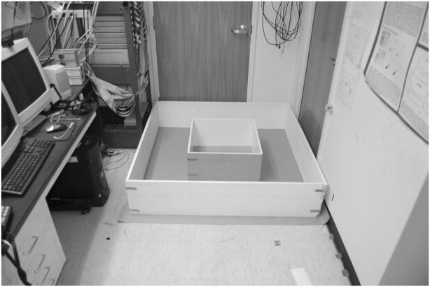
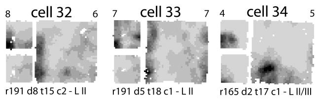
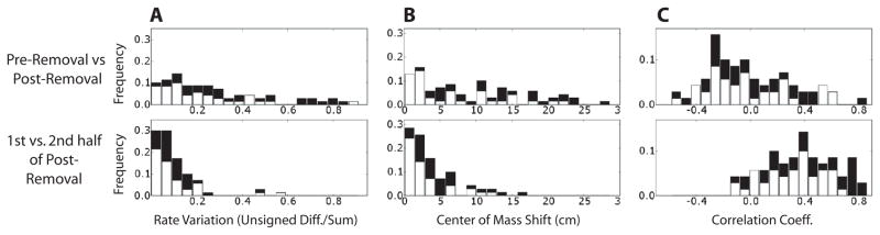
The medial entorhinal cortex (MEC) is thought to create and update a dynamical representation of the animal's spatial location. Most suggestive of this process are grid cells, whose firing locations occur periodically in space. Prior studies in small environments were ambiguous as to whether all spatially modulated cells in MEC were variants of grid cells or whether a subset resembled classic place cells of the hippocampus. Recordings from the dorsal and ventral MEC were performed as four rats foraged in a small square box centered inside a larger one. After 6 min, without removing the rat from the enclosure, the walls of the small box were quickly removed, leaving the rat free to continue foraging in the whole area enclosed by the larger box. The rate-responses of most recorded cells (70 out of 93 cells, including 15 of 16 putative interneurons) were considered spatially modulated based on information-theoretic analysis. A number of cells that resembled classic hippocampal place cells in the small box were revealed to be grid cells in the larger box. In contrast, other cells that fired along the boundaries or corners of the small box did not show grid-cell firing in the large box, but instead fired along the corresponding locations of the large box. Remapping of the spatial response in the area corresponding to the small box after the removal of its walls was prominent in most spatially modulated cells. These results show that manipulation of local boundaries can exert a powerful influence on the spatial firing patterns of MEC cells even when the manipulations leave global cues unchanged and allow uninterrupted, self-motion-based localization. Further, they suggest the presence of landmark-related information in MEC, which might prevent cumulative drift of the spatial representation or might reset it to a previously learned configuration in a familiar environment.


Figures

Figure 1
Figure 1. Recording apparatus
The foraging session was initiated in the small box centered inside the large box. The small box was composed of two L-shaped walls, which allowed its rapid removal after the rat explored the small box for approximately 6 min (PRE epoch). The rat was then free to continue foraging in the large box for the remainder of the session (POST epoch, approximately 34 min).
Figure 2
Figure 2. Representative tetrode tracks from MEC recordings (rat 191)
The dots indicate the recording sites where cells with boundary-related activity were recorded. (A) The three black filled circles indicate the progressively ventral recording locations of tetrode 17 on days 6, 7, and 8 (from the top), where cells 15, 16, 18, 19, 25, and 26 (see Fig. 4) were recorded. (B) Position of tetrode 18 on day 5, where cell 17 was recorded. (C) Position of tetrode 3 on day 3, where cell 24 was recorded. (D) Position of tetrode 1 on day 3, where cell 23 was recorded. Scale bar = 1 mm.
Figure 3
Figure 3. Spatial modulation of putative principal cells and interneurons
(A) Scatter plot of mean firing rate (m.f.r.) and spatial information (SI) of all 93 cells. The m.f.r. refers to the epoch of the session that occurred in the entire large box after the removal of the small-box walls. Filled circles denote cells (n = 70) that had a SI greater than 3 times the maximum SI computed with 100 random shufflings of the cell’s spike train and the rat’s location (see Methods). Open circles denote the cells that did not pass this test and were excluded from the remapping analysis (n = 23). Note that the two populations are respectively above and below ~0.025 bits, even though the spurious information was estimated individually for each cell. (B) Scatter plot of the m.f.r. and spike width of the 70 cells that are represented by filled circles in A. Two distinct clusters represent putative principal cells (m.f.r. < 10 Hz) and putative interneurons (m.f.r. >10 Hz).
Figure 4
Figure 4. Firing rate maps of all putative principal cells (m.f.r. < 10 Hz) with SI > 0.05 bits (see Fig. 3)
For each cell, the small map at upper left represents the small box session before the walls were removed (PRE epoch). The large map represents the large-box session after the small-box walls were removed (POST epoch). The small map at the lower left is derived from the large map by including only the pixels covered during the PRE epoch. The same grayscale is shared by all three rate maps of each cell: white denotes unvisited bins, darker shades of gray represent higher firing rates, and black is the highest of the PRE and POST peak rates (top left and right numbers respectively). Recording information is at the bottom: rat (r), day (d), tetrode (t), unit (c), and MEC layer (L II/III means a precise determination was not possible and L D denotes deep layers). Note that most of the cells shifted their fields in the small-box area after the removal of its delimiting walls.
Figure 5
Figure 5. Additional examples of putative boundary cells and an ambiguous cell
These cells had spatial information scores in the large box < 0.05, so they were not included in Figure 4. However, these cells were among the top 8 in terms of their correlation coefficients between the small box and the rescaled large box. Cells 32 and 33 fired along a wall in both boxes. Cell 34, recorded from a ventral location (Table 1), might have shown a simple grid vertex that either scaled with the environment or just revealed more of the vertex when the small box walls were removed.
Figure 6
Figure 6. Changes in spatial firing patterns and firing rates between the small and large box sessions
All spatially modulated cells were included in the three analyses. Top histograms refer to the change of response between the PRE and POST epochs (before and after the removal of the small box walls). Bottom histograms refer the change between the first and second half of the POST epoch. In both cases only the area corresponding to the small box was considered. The black components of histogram bars represent the subpopulation of putative principal cells with high spatial information (SI > 0.05 bits, all the cells displayed in Fig. 4). (A) Population distribution of the rate-change index. (B) Population distribution of the shift of the COM of the firing rate maps. (C) Population distribution of the Pearson correlation between the firing rate maps.

References

    1. Alonso A, Garcia-Austt E. Neuronal sources of theta rhythm in the entorhinal cortex of the rat. I. Laminar distribution of theta field potentials. Exp Brain Res. 1987;67:493–501. - PubMed
    1. Barnes CA, McNaughton BL, Mizumori SJ, Leonard BW, Lin LH. Comparison of spatial and temporal characteristics of neuronal activity in sequential stages of hippocampal processing. Prog Brain Res. 1990;83:287–300. - PubMed
    1. Barry C, Hayman R, Burgess N, Jeffery KJ. Experience-dependent rescaling of entorhinal grids. Nat Neurosci. 2007;10:682–684. - PubMed
    1. Barry C, Lever C, Hayman R, Hartley T, Burton S, O’Keefe J, Jeffery K, Burgess N. The boundary vector cell model of place cell firing and spatial memory. Rev Neurosci. 2006;17:71–97. - PMC - PubMed
    1. Blair HT, Kishan G, Zhang K. Conversion of a phase-coded to rate-coded position signal by a three-stage model of theta cells, grid cells, and place cells. Hippocampus. 2008;18:xxx. - PMC - PubMed

Publication types

MeSH terms