Near infrared fluorescent optical imaging for nodal staging
- PMID: 19021320
- PMCID: PMC2914597
- DOI: 10.1117/1.2953498
Near infrared fluorescent optical imaging for nodal staging
Abstract
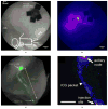
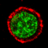
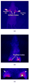
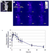
Current techniques to assess lymph node metastases in cancer patients include lymphoscintigraphy after administration of a nonspecific radiocolloid in order to locate and resect lymph nodes for pathological examination of harbored cancer cells. Clinical trials involving intradermal or subcutaneous injection of antibody-based nuclear imaging agents have demonstrated the feasibility for target-specific, molecular imaging of cancer-positive lymph nodes. The basis for employing near-infrared (NIR) optical imaging for assessing disease is evidenced by recent work showing functional lymph imaging in mice, swine, and humans. We review antibody-based immunolymphoscintigraphy with an emphasis on the use of trastuzumab (or Herceptin) to target human epidermal growth factor receptor-2 (HER2) overexpressed in some breast cancers. Specifically, we review in vitro and preclinical imaging data from our laboratory that show how the dual-labeled agent ((111)In-DTPA)(n)-trastuzumab-(IRDye800)(m) utilizes the high photon count provided by an NIR fluorescent dye, IRDye 800CW, and the radioactive signal from a gamma emitter, Indium-111, for possible detection of HER2 metastasis in lymph nodes. We show that the accumulation and clearance of ((111)In-DTPA)(n)-trastuzumab-(IRDye800)(m) from the axillary nodes of mice occurs 48 h after intradermal injection into the dorsal aspect of the foot. The requirement for long clearance times from normal, cancer-negative nodes presents challenges for nuclear imaging agents with limited half-lives but does not hamper NIR optical imaging.
Figures


References
-
- Weinstein JN, Parker RJ, Keenan AM, Dower SK, Morse HC, III, Sieber SM. Monoclonal anitbodies in the lymphatics: toward the diagnosis and therapy of tumor metastases. Science. 1982;218(4579):1334–1337. - PubMed
-
- Weinstein JN, Steller MA, Keenan AM, Covell DG, Key ME, Sieber SM, Oldham RK, Hwang KM, Parker RJ. Monoclonal antibodies in the lymphatics: selective delivery to lymph node metastases of a solid tumor. Science. 1983;222(4622):423–426. - PubMed
-
- Steller MA, Parker RJ, Covell DG, Holton OD, III, Keenan AM, Sieber SM, Weinstein JN. Optimization of monoclonal antibody delivery via the lymphatics: the dose dependence. Cancer Res. 1986;46(4 Pt 1):1830–1834. - PubMed
-
- Wahl RL, Geatti O, Liebert M, Wilson B, Shreve P, Beers BA. Kinetics of interstitially administered monoclonal antibodies for purposes of lymphoscintigraphy. J Nucl Med. 1987;28(11):1736–1744. - PubMed
-
- Mulshine JL, Keenan AM, Carrasquillo JA, Walsh T, Linnoila RI, Holton OD, Harwell J, Larson SM, Bunn PA, Weinstein JN. Immunolymphoscintigraphy of pulmonary and mediastinal lymph nodes in dogs: a new approach to lung cancer imaging. Cancer Res. 1987;47(13):3572–3576. - PubMed
Publication types
MeSH terms
Substances
Grants and funding
LinkOut - more resources
Full Text Sources
Other Literature Sources
Medical
Research Materials
Miscellaneous

