Review
. 2009 May;11(5):1029-46.
doi: 10.1089/ars.2008.2296.

Iron-based redox switches in biology

Affiliations
Review

Iron-based redox switches in biology

F Wayne Outten et al. Antioxid Redox Signal. 2009 May.

Abstract

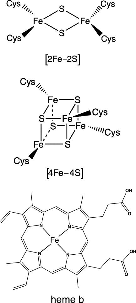
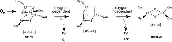
By virtue of its unique electrochemical properties, iron makes an ideal redox active cofactor for many biologic processes. In addition to its important role in respiration, central metabolism, nitrogen fixation, and photosynthesis, iron also is used as a sensor of cellular redox status. Iron-based sensors incorporate Fe-S clusters, heme, and mononuclear iron sites to act as switches to control protein activity in response to changes in cellular redox balance. Here we provide an overview of iron-based redox sensor proteins, in both prokaryotes and eukaryotes, that have been characterized at the biochemical level. Although this review emphasizes redox sensors containing Fe-S clusters, proteins that use heme or novel iron sites also are discussed.
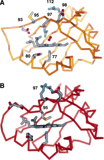

Figures

FIG. 1.
FIG. 1.
Iron cofactors commonly used in biology. From top to bottom, heme b, [2Fe-2S] cluster, [4Fe-4S] cluster, and heme b.
FIG. 2.
FIG. 2.
(A) Crystal structure of oxidized [2Fe-2S] SoxR dimer bound to DNA (stick representation). For SoxR, blue is the DNA-binding domain, magenta is the dimerization helix, and yellow is the Fe-S cluster-binding domain with red spheres as iron and green spheres as sulfur. (B) A close-up showing the interactions between the Fe-S cluster-binding domain of one SoxR monomer and the DNA-binding domain of the other monomer (shown in white for clarity). (Reproduced with permission from ref. 160). (For interpretation of the references to color in this figure legend, the reader is referred to the web version of this article at www.liebertonline.com/ars).
FIG. 3.
FIG. 3.
(A) Protein environment surrounding the [2Fe-2S]2+ cluster of SoxR when bound to DNA. Iron and sulfur are red and green spheres, and NH-S hydrogen bonds are dashed lines. (B) Proposed model of electrostatic changes that occur around the SoxR [2Fe-2S] cluster on reduction (addition of e-). (Reproduced with permission from ref. 160). (For interpretation of the references to color in this figure legend, the reader is referred to the web version of this article at www.liebertonline.com/ars).
FIG. 4.
FIG. 4.
[4Fe-4S] cluster oxidation and disassembly on exposure of FNR to oxygen (see text for details).
FIG. 5.
FIG. 5.
A comparison of the structure of the polypeptide IRP1 bound to [4Fe-4S] (c-aconitase) on the left and bound to the IRE-RNA, noncoding regulatory structure on the right. Note the large difference in structure, indicating the possibility that the redox- sensitive “switch” occurs at or before [4Fe-4S]2+ transfer from the CIA cytosolic Fe-S assembly machinery to the apo-IRP1 polypeptide. (Reproduced with permission from ref. 158). (For interpretation of the references to color in this figure legend, the reader is referred to the web version of this article at www.liebertonline.com/ars).
FIG. 6.
FIG. 6.
Crystal structure of EcDOS heme showing rearrangments in heme pocket on oxygen binding. (A) EcDOS-O2 (yellow) with His77 (below the heme plane) in the proximal position and O2 as two red spheres above the heme. (B) Deoxygenated EcDOS (red). Amino acids are numbered according to EcDOS sequence. (Reproduced with permission from ref. 111). (For interpretation of the references to color in this figure legend, the reader is referred to the web version of this article at www.liebertonline.com/ars).

Similar articles

Cited by

References

    1. Agron PG. Ditta GS. Helinski DR. Oxygen regulation of nifA transcription in vitro. Proc Natl Acad Sci U S A. 1993;90:3506–3510. - PMC - PubMed
    1. Alen C. Sonenshein AL. Bacillus subtilis aconitase is an RNA-binding protein. Proc Natl Acad Sci U S A. 1999;96:10412–10417. - PMC - PubMed
    1. Althaus EW. Outten CE. Olson KE. Cao H. O'Halloran TV. The ferric uptake regulation (Fur) repressor is a zinc metalloprotein. Biochemistry. 1999;38:6559–6569. - PubMed
    1. Andrews SC. Robinson AK. Rodriguez-Quinones F. Bacterial iron homeostasis. FEMS Microbiol Rev. 2003;27:215–237. - PubMed
    1. Anxolabehere-Mallart E. Glaser T. Frank P. Aliverti A. Zanetti G. Hedman B. Hodgson KO. Solomon EI. Sulfur K-edge X-ray absorption spectroscopy of 2Fe-2S ferredoxin: covalency of the oxidized and reduced 2Fe forms and comparison to model complexes. J Am Chem Soc. 2001;123:5444–5452. - PubMed

Publication types