. 2010 Jan;20(1):66-79.
doi: 10.1111/j.1750-3639.2008.00244.x. Epub 2008 Nov 19.

Modeling the association between 43 different clinical and pathological variables and the severity of cognitive impairment in a large autopsy cohort of elderly persons

Affiliations

Modeling the association between 43 different clinical and pathological variables and the severity of cognitive impairment in a large autopsy cohort of elderly persons

Peter T Nelson et al. Brain Pathol. 2010 Jan.

Abstract

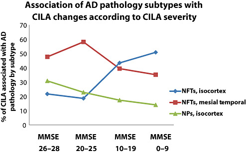
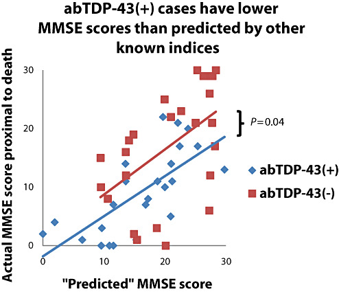
We evaluated the association between mini-mental status examination (MMSE) scores proximal to death and the values of 43 different clinical and pathological parameters. Studies were performed using data from 334 elderly, longitudinally evaluated research subjects who had undergone autopsy and satisfied inclusion criteria from an initial study group of 501. Interindividual variance in MMSE scores was used as a surrogate for the severity of cognitive impairment linked to aging (CILA). A statistical linear regression-based model provided a framework for assessing the parameters with significant, direct impact on CILA severity. Strong association between CILA and Alzheimer's disease (AD) pathology, especially isocortical neurofibrillary tangles, was evident. The pattern of association between AD lesion densities with cognitive impairment severity was biologically informative, with neuritic plaques having more impact in relatively high-functioning individuals. Abundant isocortical Lewy bodies tended to be an additive pathology correlating with final MMSE scores approximately 10 points lower. In a subset of cases we found evidence for association between TDP-43-related pathology and CILA severity, independent of AD or hippocampal sclerosis. There was no support for independent association between CILA severity and most evaluated indices including diffuse plaques, argyrophilic grains, heart disease, education level, apolipoprotein E alleles or diabetes.


Figures

Figure 1
Figure 1
The association of particular subtypes and anatomical areas of Alzheimer's disease (AD) pathology with cognitive impairment linked to aging (CILA) severity changes according to the different stages of cognitive impairment. This chart shows data from modeling on the entire cohort using the statistically significant parameter coefficients as shown in Table 5. Cases are binned by mini‐mental status examination (MMSE) scores: MMSE 26–28 (n = 83); MMSE 20–26 (n = 38); MMSE 10–20 (n = 65); and MMSE 0–10 (n = 62). Note that as cognition declines, the apparent impact of isocortical neurofibrillary tangles (NFTs) becomes greater, and the relative importance of mesial temporal lobe NFTs and isocortical NPs declines. Nonetheless, each subtype of AD‐type pathology is associated independently with significant amount of CILA.
Figure 2
Figure 2
Statistical methods indicated that argyrophilic grains (AGs) are not associated with CILA severity in our research cohorts. To evaluate these data, we studied separately the 75 cases with AGs in our sample. A chart (A) shows how the actual pre‐mortem mini‐mental status examination (MMSE) scores for patients correlated to the amount of AG pathology. The AG score is proportional to the severity of AGs in entorhinal cortex, subiculum and cornu ammonis hippocampal fields. Note that there is no decrease in MMSE scores as AG pathology worsens. A photomicrograph of Gallyas‐stained cornu ammonis (B) from the individual in our autopsy cohort with the most severe AG pathology, a 91‐year old woman who died without any cognitive impairment (MMSE score = 30 less than 8 months prior to death). Scale bar = 50 µm.
Figure 3
Figure 3
The multiple variable linear regression algorithm allows the prediction of mini‐mental status examination (MMSE) for each case based on the parameter coefficients. The algorithm was applied using the parameter coefficients from the combined cohort as shown in Table 5 for the 49 cases stained for TDP‐43 immunohistochemistry. The results indicate that the abTDP‐43(+) show a significant downward shift such that the “actual” MMSE score was lower than the abTDP‐43(−) cases (difference of intercept P = 0.04), indicating that abTDP‐43 is associated with cognitive impairment linked to aging severity independent of other known risk factors such as Alzheimer's disease or hippocampal sclerosis pathology.
Figure 4
Figure 4
Photomicrographs of TDP‐43 immunohistochemistry (IHC) show abnormal (A,C) and normal (B,D) staining, which can be a challenge to discriminate. Unlike most neurodegeneration‐related immunostains, TDP‐43 IHC is strongly positive in normal tissues. In some end‐stage AD cases we interpreted to be abTDP‐43(−) there were “regularly irregular” staining in the dentate granule cells (B) and hyperchromatic cells (D, horizontal arrows) that were different from the normal‐appearing cells (vertical arrow) but distinct also from the aberrant intracellular inclusions (C). AbTDP‐43 staining in CA1 (C) showing the neurofibrillary tangle‐like (arrow) and neuritic thread‐like structures (arrows). Immunopositivity was scored blindly. Scale bars = 30 µm (A,B) or 50 µm (C,D).
Figure 5
Figure 5
A theoretical model that harmonizes data from the present study and prior clinico‐pathological correlation research. Because isocortical neuritic plaques (NPs) and hippocampal neurofibrillary tangles (NFTs) have significant “floor” and “ceiling” effects in relationship to the course of dementia, they are not strongly related to the cognitive impairment linked to aging (CILA) severity prior to autopsy (87). However, they may still play a strong pathogenetic role and contribute directly to CILA particularly in the earliest stages of Alzheimer's disease.

Similar articles

Cited by

References

    1. Akram A, Christoffel D, Rocher AB, Bouras C, Kovari E, Perl DP et al (2008) Stereologic estimates of total spinophilin‐immunoreactive spine number in area 9 and the CA1 field: relationship with the progression of Alzheimer's disease. Neurobiol Aging 29:1296–1307. - PMC - PubMed
    1. Amador‐Ortiz C, Ahmed Z, Zehr C, Dickson DW (2007) Hippocampal sclerosis dementia differs from hippocampal sclerosis in frontal lobe degeneration. Acta Neuropathol (Berl) 113:245–252. - PMC - PubMed
    1. Amador‐Ortiz C, Lin WL, Ahmed Z, Personett D, Davies P, Duara R et al (2007) TDP‐43 immunoreactivity in hippocampal sclerosis and Alzheimer's disease. Ann Neurol 61:435–445. - PMC - PubMed
    1. Arai T, Hasegawa M, Akiyama H, Ikeda K, Nonaka T, Mori H et al (2006) TDP‐43 is a component of ubiquitin‐positive tau‐negative inclusions in frontotemporal lobar degeneration and amyotrophic lateral sclerosis. Biochem Biophys Res Commun 351:602–611. - PubMed
    1. Armstrong RA (2003) Quantifying the pathology of neurodegenerative disorders: quantitative measurements, sampling strategies and data analysis. Histopathology 42:521–529. - PubMed

Publication types

MeSH terms