. 2008 Nov 20:9:552.
doi: 10.1186/1471-2164-9-552.

Exploring the mialome of ticks: an annotated catalogue of midgut transcripts from the hard tick, Dermacentor variabilis (Acari: Ixodidae)

Affiliations

Exploring the mialome of ticks: an annotated catalogue of midgut transcripts from the hard tick, Dermacentor variabilis (Acari: Ixodidae)

Jennifer M Anderson et al. BMC Genomics. .

Abstract

Background: Ticks are obligate blood feeders. The midgut is the first major region of the body where blood and microbes ingested with the blood meal come in contact with the tick's internal tissues. Little is known about protein expression in the digestive tract of ticks. In this study, for analysis of global gene expression during tick attachment and feeding, we generated and sequenced 1,679 random transcripts (ESTs) from cDNA libraries from the midguts of female ticks at varying stages of feeding.

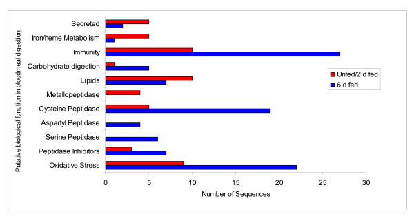
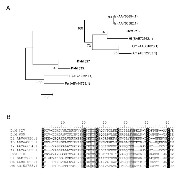
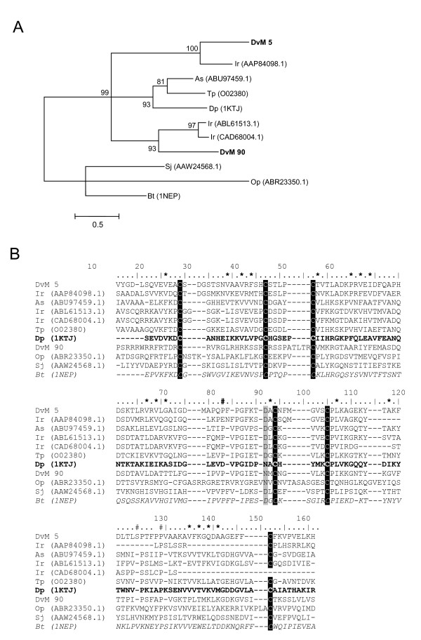
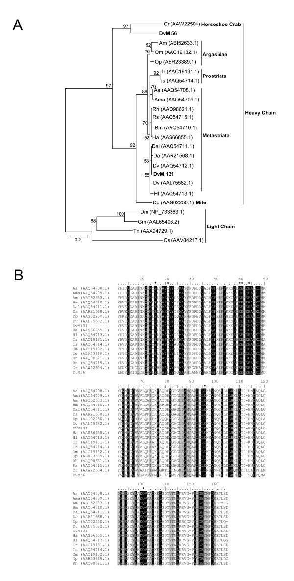
Results: Sequence analysis of the 1,679 ESTs resulted in the identification of 835 distinct transcripts, from these, a total of 82 transcripts were identified as proteins putatively directly involved in blood meal digestion, including enzymes involved in oxidative stress reduction/antimicrobial activity/detoxification, peptidase inhibitors, protein digestion (cysteine-, aspartic-, serine-, and metallo-peptidases), cell, protein and lipid binding including mucins and iron/heme metabolism and transport. A lectin-like protein with a high match to lectins in other tick species, allergen-like proteins and surface antigens important in pathogen recognition and/or antimicrobial activity were also found. Furthermore, midguts collected from the 6-day-fed ticks expressed twice as many transcripts involved in bloodmeal processing as midguts from unfed/2-day-fed ticks.

Conclusion: This tissue-specific transcriptome analysis provides an opportunity to examine the global expression of transcripts in the tick midgut and to compare the gut response to host attachment versus blood feeding and digestion. In contrast to those in salivary glands of other Ixodid ticks, most proteins in the D. variabilis midgut cDNA library were intracellular. Of the total ESTs associated with a function, an unusually large number of transcripts were associated with peptidases, cell, lipid and protein binding, and oxidative stress or detoxification. Presumably, this is consistent with their role in intracellular processing of the blood meal and response to microbial infections. The presence of many proteins with similar functions is consistent with the hypothesis that gene duplication contributed to the successful adaptation of ticks to hematophagy. Furthermore, these transcripts may be useful to scientists investigating the role of the tick midgut in blood-meal digestion, antimicrobial activity or the transmission of tick-borne pathogens.


Figures

Figure 1
Figure 1
Differential display of proteins associated with midgut function either unfed and 2 days post bloodmeal (unfed/2 d fed) or 6 days post bloodmeal (6 d fed).
Figure 2
Figure 2
SDS-PAGE protein gel from a lysate of midguts from 6 d-fed female Dermacentor variabilis showing the location of proteins identified by tryptic digestion-mass spectrometry. Abbreviations: Fr 25, Fr 26 = fractions 25 and 26; Glyphosph = Glycogen phosphorylase; G-3-pdh = glyceraldehydes-3-phosphodehydrogenase; GST = Glutathione S-transferase; HPLC = high performance liquid chromatography; HSP = 70 kD heat shock protein; Mem. protein = Bm86 membrane antigen in Rhipicephalus (Boophilus) microplus; MW = molecular weight markers; PC-oxid = prenylcysteine oxidase. Balanus amphrite (Ba), Rhipicephalus (Boophilus) microplus (Bm), D. variabilis (Dv), Homo sapiens (Hs), Ixodes pacificus (Ip), Pleocyemata unclassified (Pl), Rattus norvegicus (Rn) and Xenopus tropicalis (Xt).
Figure 3
Figure 3
Analysis of Glutathione-S-transferase (GST) protein family. Phylogenetic tree based on maximum likelihood analysis of Dermacentor variabilis midgut protein and published GST sequences. Transcripts identified in this analysis are in bold. Phylogenetic analysis was conducted on protein alignments using Tree Puzzle version 5.2. Values at the nodes represent calculated internal branch node support (1000 replications). D. variabilis (Dv), Ixodes pacificus (Ip), Sarcopte scabiei (Ss), Araneus ventricosus (Av), Rhipicephalus appendiculatus (Ra), R. (Boophilus) microplus (Bm), Haemaphysalis longicornis (Hl), Dermatophagoides pteronyssinus (Dp), and Psoroptes ovis (Po).
Figure 4
Figure 4
Multiple sequence alignment of protein sequences identified in a cDNA library of unfed/2 d fed or 6 d fed D. variabilis midguts (DvM) and published tick GST sequences found on genbank. Catalytic residues are indicated by an asterisk and conserved residues involved in the GST fold are indicated by arrowheads. The conserved SMAIL domain is boxed. Shading represents 100% identity (black) or similarity (grey) among the sequences. Alignments were conducted using CLUSTALX. D. variabilis (Dv), Ixodes pacificus (Ip), Rhipicephalus appendiculatus (Ra), R. (Boophilus) microplus (Bm), and Haemaphysalis longicornis (Hl).
Figure 5
Figure 5
Analysis of microsomal GST protein sequences. (A) Phylogenetic tree based on maximum likelihood analysis Dermacentor variabilis midgut protein and published microsomal GST sequences. The transcript identified in this analysis is in bold (DvM). Phylogenetic analysis was conducted on protein alignments using Tree Puzzle version 5.2. Values at nodes represent calculated internal branch node support (1000 replications). (B) Multiple sequence alignment (CLUSTALX) of protein sequences identified in a cDNA library of unfed/2 d fed or 6 d fed D. variabilis midguts (DvM) and published microsomal GST 3 sequences found on genbank. Shading represents 100% identity (black) or similarity (grey) among the sequences. Alignments were conducted using CLUSTALX. D. variabilis (Dv), Rattus norvegicus (Nr), Mus musculus (Mm), Canis familiaris (Cf), Xenopus tropicalis (Xt), Strongylocentrotus purpuratus (Sp), Escherichia coli (Ec), Yersinia pestis (Yp), Homo sapiens (Hs), Bordetella pertussis (Bp), Prochlorococcus marinus (Pm), Drosophila melanogaster (Dm), Anopheles gambiae (Ag).
Figure 6
Figure 6
Analysis of Thioredoxin (TRX) protein family. (A) Phylogenetic tree based on maximum likelihood analysis of a Dermacentor variabilis midgut protein and published TRX sequences. The transcript identified in this analysis is in bold (DvM). Phylogenetic analysis was conducted on protein alignments using Tree Puzzle version 5.2. Values at nodes represent calculated internal branch node support (1000 replications). (B) Multiple sequence alignment (CLUSTALX) of protein sequences identified in a cDNA library of unfed/2 d fed or 6 d fed D. variabilis midguts (DvM) and published thiroredoxin-1 sequences found on genbank. Arrowheads indicate the catalytic cysteine motif. Shading represents 100% identity (black) or similarity (grey) among the sequences. Alignments were conducted using CLUSTALX. D. variabilis (Dv), Ixodes scapularis (Is), Mesobuthus cyprius (Mc), Aedes aegypti (As), Anopheles gambiae (Ag), Drosophila melanogaster (Dm), Bombyx mori (Bm), Manduca sexta (Ms), Tribolium castaneum (Tc), Maconellicoccus hirsutus (Mh), Drosophila pseudoobscura (Dp), Simulium jonesii (Sj), and Homo sapiens (Hs).
Figure 7
Figure 7
Analysis of Glutaredoxin (GRX) protein family. Phylogenetic tree based on maximum likelihood analysis of a Dermacentor. variabilis midgut protein and published glutaredoxin (GRX) sequences. The transcript identified in this analysis is in bold (DvM). Phylogenetic analysis was conducted on protein alignments using Tree Puzzle version 5.2. Values at nodes represent calculated internal branch node support (1000 replications). D. variabilis (Dv), Plasmodium falciparum (Pf), Drosophila melanogaster (Dm), Anopheles gambiae (Ag), Bombyx mori (Bm), Tribolium castaneum (Tc), Aedes aegypti (Aa).
Figure 8
Figure 8
Analysis of Phospholipid-Hydroperoxide Glutathione Peroxidase (PHGPx) protein family. Multiple sequence alignment (CLUSTALX) of protein sequences identified in a cDNA library of unfed/2 d fed or 6 d fed Dermacentor. variabilis midguts (DvM) and Rhipicephalus (Boophilus) microplus (Bm) PHGPx found on genbank. Arrowhead indicates the position of the selenocysteine (X) and the diamond (◆) indicates the active-site residues Gln and Trp that interact with the selenocysteine. Shading represents 100% identity (black) or similarity (grey) among the sequences. Alignments were conducted using CLUSTALX.
Figure 9
Figure 9
Analysis of Cu, Zn Superoxide dismutase (SOD) protein family. Multiple sequence alignment (CLUSTALX) of protein sequences identified in a cDNA library of unfed/2 d fed or 6 d fed Dermacentor variabilis midguts (DvM), Argas monolakensis (Am) and Ixodes scapularis (Is) Cu, Zu SOD. Arrowhead indicates the position of residues that bind either copper (C) or zinc (Z) and the residues that form the disulfide bridges (S-S). Shadding respresents 100% identity (black) or similarity (grey). Alignments were conducted using CLUSTALX.
Figure 10
Figure 10
Analysis of serine peptidase inhibitors (serpins). Phylogenetic tree based on maximum likelihood analysis Dermacentor variabilis midgut protein and published serpin sequences from various tick species including 17 serpins, known as lospin, from Amblyomma americanum. The transcript identified in this analysis is in bold (DvM). Phylogenetic analysis was conducted on protein alignments using Tree Puzzle version 5.2. Values at nodes represent calculated internal branch node support (1000 replications). D. variabilis (Dv), Ixodes scapularis (Is), Amblyomma americanum (Aa), Haemaphysalis longicornis (Hl), Rhipicephalus appendiculatus (Ra), I. ricinus (Ir), Drosophila melanogaster (Dm), R. (Boophilus) microplus (Bm), Brugia malayi (Bma).
Figure 11
Figure 11
Analysis of Boophilin-like peptidase inhibitors. Multiple sequence alignment (CLUSTALX) of protein sequences identified in a cDNA library of unfed/2 d fed or 6 d fed Dermacentor variabilis midguts (DvM) and thrombin in hibitor-like proteins found on genbank. Black shading indicates identity and grey shading indicates similarity. Boxed area indicates the amino acids that constitute the peptidase recognition loop. * P1 site, the primary recognition residue. Arrowhead indicates the start of the second Kunitz domain. Shading represents 100% identity (black) or similarity (grey) among the sequences. Alignments were conducted using CLUSTALX. D. variabilis (Dv), Homo sapiens alpha-1-microglobulin bikunin (Hs), Pancreatic trypsin inhibitor precursor (BPTI), Amblyomma hebraeum (Ah), Rhipicephalus (Boophilus) microplus (Bm), Ixodes scapularis (Is), Ornithodoros moubata (Om), O. savignyi (Os).
Figure 12
Figure 12
Analysis of Cystatin a family of cystine peptidases. (A) Phylogenetic tree based on maximum likelihood analysis of a Dermacentor variabilis midgut protein and published secreted and cytoplasmic cystatin sequences. The transcripts identified in this analysis are in bold (DvM). Phylogenetic analysis was conducted on protein alignments using Tree Puzzle version 5.2. Values at nodes represent calculated internal branch node support (1000 replications). Multiple sequence alignment (CLUSTALX) of protein sequences identified in a cDNA library of unfed/2 d fed or 6 d fed D. variabilis midguts (DvM) and published secreted (B) and cytoplasmic (C) cystatin sequences found on Genbank. Arrowheads indicate putative papain binding domains. S-S indicates cysteines involved in disulfide bridge formation. The underlined amino acids highlights the conserved QNVLG domain. Shading represents 100% identity (black) or similarity (grey) among the sequences. Alignments were conducted using CLUSTALX.D. variabilis (Dv), Ixodes scapularis (Is), I. ricinus (Ir), Ornithodoros moubata (Om), Haemaphysalis longicornis (Hl), Dictyostelium discoideum (Dd), and Lepidoglyphus destructor (Ld).
Figure 13
Figure 13
Analysis of serine peptidases. Multiple sequence alignment (CLUSTALX) of protein sequences identified in a cDNA library of unfed/2 d fed or 6 d fed Dermacentor variabilis midguts (DvM) and tick serine peptidases found on genbank. Arrowheads conserved amino acids involved in the catalytic triad. Shading represents 100% identity (black) or similarity (grey) among the sequences. Alignments were conducted using CLUSTALX. D. variabilis (Dv), Ornithodoros moubata (Om), Haemaphysalis longicornis (Hl), Rhipicephalus appendiculatus (Ra).
Figure 14
Figure 14
Analysis of aspartic peptidases, Cathepsin D. (A) Phylogenetic tree based on maximum likelihood analysis of a Dermacentor variabilis midgut protein and published sequences. The transcripts identified in this analysis are in bold (DvM). Phylogenetic analysis was conducted on protein alignments using Tree Puzzle version 5.2. Values at nodes represent calculated internal branch node support (1000 replications). (B) Multiple sequence alignment (CLUSTALX) of protein sequences identified in a cDNA library of unfed/2 d fed or 6 d fed D. variabilis midguts (DvM) and published sequences found on Genbank. Arrowheads indicate conserved aspartic catalytic sites and diamonds indicate putative glycosylation residues (nomenclature derived from Boldaatar et al. (49). Shading represents 100% identity (black) or similarity (grey) among the sequences. Alignments were conducted using CLUSTALX. D. variabilis (Dv), Drosophila melanogaster (Dm), Bombyx mori (Bmori), Haemaphysalis longicornis (Hl), Ixodes ricnius (Ir), and Rhipicephalus (Boophilus) microplus (Bm), Ancylostoma caninum (Ac), Sus scrofa (Ss), Rattus norvegicus (Rn).
Figure 15
Figure 15
Analysis of aspartic endopeptidases (AE), a legumain-like family of proteins. (A) Phylogenetic tree based on maximum likelihood analysis of a Dermacentor variabilis midgut protein and published legumain-like sequences. The transcript identified in this analysis is in bold (DvM). Phylogenetic analysis was conducted on protein alignments using Tree Puzzle version 5.2. Values at nodes represent calculated internal branch node support (1000 replications). (B) Multiple sequence alignment (CLUSTALX) of protein sequences identified in a cDNA library of unfed/2 d fed or 6 d fed D. variabilis midguts (DvM) and published sequences from other ticks found on Genbank. Arrowheads indicate conserved His and Cys residues forming the catalytic dyad of AE. Underlined amino acids represent the predicted cleavage position of the signal peptide. Shading represents 100% identity (black) or similarity (grey) among the sequences. Alignments were conducted using CLUSTALX. D. variabilis (Dv), Homo sapiens (Hs), Mus musculus (Mm), Xenopus laevis (Xl), Xenopus tropicalis (Xt), Haemaphysalis longicornis (Hl), Ixodes ricnius (Ir), Schistosoma mansoni (Sm), Schistosoma japonicum (Sj), Fasciola hepatica (Fh), Caenorhabditis elegans (Ce), Arabidopsis thaliana (At), and Oryza sativa (Os).
Figure 16
Figure 16
Analysis of cysteine peptidases. (A) Phylogenetic tree based on maximum likelihood analysis of a Dermacentor variabilis midgut protein and published tick Cathepsin L and B-like peptidase sequences. The transcript identified in this analysis is in bold (DvM). Phylogenetic analysis was conducted on protein alignments using Tree Puzzle version 5.2. Values at nodes represent calculated internal branch node support (1000 replications). (B) Multiple sequence alignment (CLUSTALX) of protein sequences identified in a cDNA library of unfed/2 d fed or 6 d fed D. variabilis midguts (DvM) and published sequences from other ticks found on Genbank. Arrowheads indicate catalytic residues. Diamonds represent predicted amino acids involved in catalysis. Shading represents 100% identity (black) or similarity (grey) among the sequences. Alignments were conducted using CLUSTALX. D. variabilis (Dv), Ixodes ricinus (Ir), Haemaphysalis longicornis (Hl), Rhipicephalus (Boophilus) microplus (Bm), R. appendiculatus (Ra), R. haemaphysaloides (Rh).
Figure 17
Figure 17
Analysis of mucins. (A) Phylogenetic tree based on maximum likelihood analysis of a Dermacentor variabilis midgut protein and published mucin-like protein sequences. The transcript identified in this analysis is in bold (DvM). Phylogenetic analysis was conducted on protein alignments using Tree Puzzle version 5.2. Values at nodes represent calculated internal branch node support (1000 replications). (B) Multiple sequence alignment (CLUSTALX) of protein sequences identified in a cDNA library of unfed/2 d fed or 6 d fed D. variabilis midguts (DvM) and published sequences found on Genbank. The conserved cysteines are highlighted. Shading represents 100% identity (black) or similarity (grey) among the sequences. Alignments were conducted using CLUSTALX. D. variabilis (Dv), Ixodes scapularis (Is), Haemaphysalis longicornis (Hl), Ornithodoros moubata (Om), Argas monolakensis (Am), Lutzomyia longipalpis (Ll), Phlebotomus papatasi (Pp).
Figure 18
Figure 18
Analysis of ML domain containing proteins. (A) Phylogenetic tree based on maximum likelihood analysis of a Dermacentor variabilis midgut protein and published ML domain protein sequences. The transcripts identified in this analysis are in bold (DvM). Phylogenetic analysis was conducted on protein alignments using Tree Puzzle version 5.2. Values at nodes represent calculated internal branch node support (1000 replications). (B) Multiple sequence alignment (CLUSTALX) of protein sequences identified in a cDNA library of unfed/2 d fed or 6 d fed D. variabilis midguts (DvM) and published sequences found on Genbank. The conserved cysteines are highlighted. Number signs (#) represents putative cholesterol/lipid binding site sites based on the conserved domain of Niemann-Pick type C2 (Npc2) proteins (italisized). Asterisks (*) indicated amino acids involved in the putative lipid binding cavity based on ML domain of the dust mite allergen, Der P 2 (bold). Shading represents 100% identity (black) or similarity (grey) among the sequences. Alignments were conducted using CLUSTALX. D. variabilis (Dv), Ixodes ricinus (Ir), Acarus siro (As), Schistosoma japonicum (Sj), Dermatophagoides pteronyssius (Dp), Ornithodoros parkeri (Op), Tyrophagus putrescentiae (Tp), Bos Taurus (Bt).
Figure 19
Figure 19
Analysis of ferritin proteins. (A) Phylogenetic tree based on maximum likelihood analysis of a Dermacentor variabilis midgut protein and published heavy and light chain ferritin protein sequences. The transcripts identified in this analysis are in bold (DvM). Phylogenetic analysis was conducted on protein alignments using Tree Puzzle version 5.2. Values at nodes represent calculated internal branch node support (1000 replications). (B) Multiple sequence alignment (CLUSTALX) of protein sequences identified in a cDNA library of unfed/2 d fed or 6 d fed D. variabilis midguts (DvM) and published heavy chain ferritins found on Genbank. Asterisks (*) represent the ferroxidase diiron center based on the ferritin conserved domain (cd00904). Shading represents 100% identity (black) or similarity (grey) among the sequences. Alignments were conducted using CLUSTALX.D. variabilis (Dv), Argas monolakensis (Am), Ornithodoros moubata (Om), O. parkeri (Op), Ixodes ricinus (Ir), I. scapularis (Is), Amblyomma americanum (Aa), Amblyomma maculatum (Ama), Rhipicephalus (Boophilus) microplus (Bm), Hyalomma asiaticum (Ha), R. haemaphysaloides (Rh), Rhipicephalus sanguineus (Rs), Dermacentor variabilis (Dv), Dermacentor albipictus (Dal), Dermacentor andersoni (Da), Haemaphysalis longicornis (Hl), Dermatophagoides pteronyssinus (Dp), Carcinoscorpius rotundicauda (Cr), Drosophila melanogaster (Dm), Culicoides sonorensis (Cs), Trichoplusia ni (Tn), Glossina morsitans (Gm).

References

    1. Fogaca AC, da Silva PI, Jr, Miranda MT, Bianchi AG, Miranda A, Ribolla PE, Daffre S. Antimicrobial activity of a bovine hemoglobin fragment in the tick Boophilus microplus. J Biol Chem. 1999;274:25330–25334. - PubMed
    1. Nakajima Y, Ogihara K, Taylor D, Yamakawa M. Antibacterial hemoglobin fragments from the midgut of the soft tick, Ornithodoros moubata (Acari: Argasidae) J Med Entomol. 2003;40:78–81. - PubMed
    1. Sonenshine DE, Hynes WL, Ceraul SM, Mitchell R, Benzine T. Host blood proteins and peptides in the midgut of the tick Dermacentor variabilis contribute to bacterial control. Exp Appl Acarol. 2005;36:207–223. - PubMed
    1. Nakajima Y, Goes van Naters-Yasui A van der, Taylor D, Yamakawa M. Antibacterial peptide defensin is involved in midgut immunity of the soft tick, Ornithodoros moubata. Insect Mol Biol. 2002;11:611–618. - PubMed
    1. Kopacek P, Vogt R, Jindrak L, Weise C, Safarik I. Purification and characterization of the lysozyme from the gut of the soft tick Ornithodoros moubata. Insect Biochem Mol Biol. 1999;29:989–997. - PubMed

Publication types

LinkOut - more resources