

Interferon regulatory factor 6 (IRF6) is expressed in the ovine uterus and functions as a transcriptional activator
- PMID: 19022341
- PMCID: PMC2655364
- DOI: 10.1016/j.mce.2008.10.025
Interferon regulatory factor 6 (IRF6) is expressed in the ovine uterus and functions as a transcriptional activator
Abstract
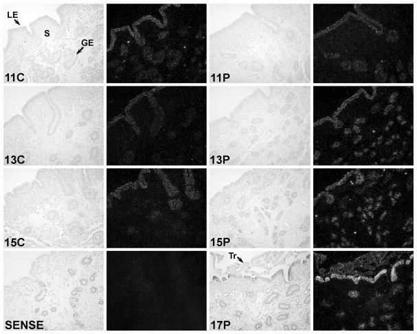
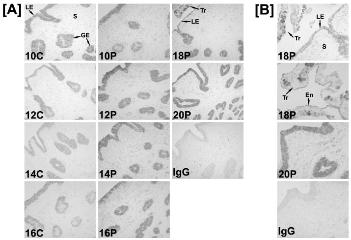
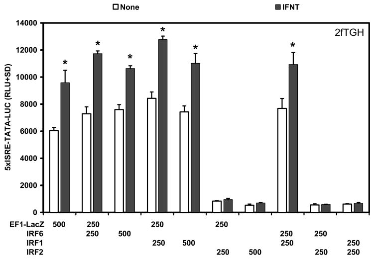
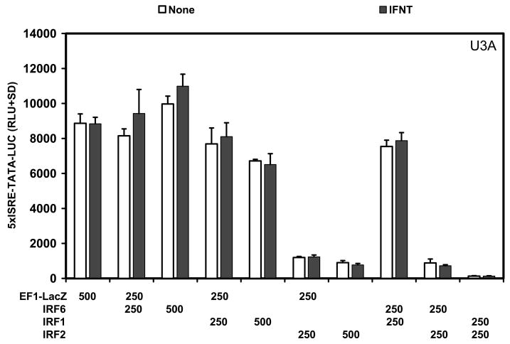
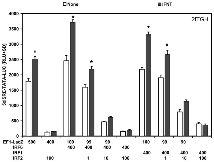
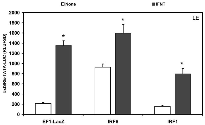
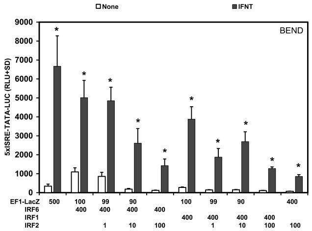
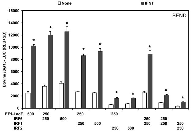
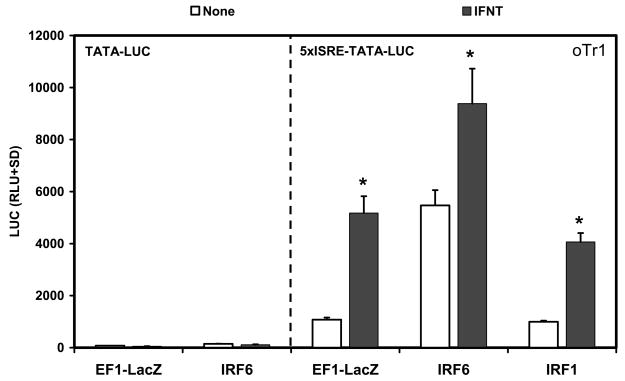
Interferon tau (IFNT), the maternal recognition of pregnancy signal in sheep and other ruminants, is secreted by the conceptus and regulates the expression of a number of genes in a cell-specific manner within the uterus. The response of different endometrial cell types to IFNT appears to be specified by IFN regulatory factors (IRFs). IRF2, a potent repressor of gene transcription, is expressed only by luminal (LE) and superficial glandular epithelia (sGE), whereas IRF1 and IRF9, activators of gene transcription, are expressed only in GE and stromal cells of the uterus during early pregnancy. In the present study, IRF6 was found to be expressed in LE/sGE and middle GE of the ovine uterine endometrium as well as conceptus trophectoderm. IRF family members can regulate transcription via IFN-stimulated response elements (ISREs). Transient transfection analyses found that IRF6 enhanced basal activity of ISRE-containing promoters, but did not enhance IFNT stimulation of ISRE-containing promoters in variety of different cell types. Further, IRF6 did not cooperate with IRF1 or reduce IRF2 repression of ISRE-containing promoter activity. These results establish that IRF6 is a transcriptional activator that is preferentially expressed in the endometrial epithelia and conceptus trophectoderm. IRF6 is hypothesized to play critical roles in endometrial gene expression as well as in conceptus trophectoderm growth and differentiation.
Figures
Similar articles
-
Interferon regulatory factor-two restricts expression of interferon-stimulated genes to the endometrial stroma and glandular epithelium of the ovine uterus.Biol Reprod. 2001 Oct;65(4):1038-49. doi: 10.1095/biolreprod65.4.1038. Biol Reprod. 2001. PMID: 11566724
-
Progesterone and interferon tau regulate leukemia inhibitory factor receptor and IL6ST in the ovine uterus during early pregnancy.Reproduction. 2009 Mar;137(3):553-65. doi: 10.1530/REP-08-0437. Epub 2008 Dec 5. Reproduction. 2009. PMID: 19060097
-
Progesterone and interferon tau regulate hypoxia-inducible factors in the endometrium of the ovine uterus.Endocrinology. 2008 Apr;149(4):1926-34. doi: 10.1210/en.2007-1530. Epub 2008 Jan 3. Endocrinology. 2008. PMID: 18174278 Free PMC article.
-
Biology of progesterone action during pregnancy recognition and maintenance of pregnancy.Front Biosci. 2002 Sep 1;7:d1879-98. doi: 10.2741/spencer. Front Biosci. 2002. PMID: 12161340 Review.
-
Interferons and progesterone for establishment and maintenance of pregnancy: interactions among novel cell signaling pathways.Reprod Biol. 2008 Nov;8(3):179-211. doi: 10.1016/s1642-431x(12)60012-6. Reprod Biol. 2008. PMID: 19092983 Review.
Cited by
-
Experimental and bioinformatic analysis of cultured Bovine Endometrial Cells (BEND) responding to interferon tau (IFNT).Reprod Biol Endocrinol. 2016 Apr 18;14:22. doi: 10.1186/s12958-016-0156-y. Reprod Biol Endocrinol. 2016. PMID: 27091464 Free PMC article.
-
Comparative analysis between endometrial proteomes of pregnant and non-pregnant ewes during the peri-implantation period.J Anim Sci Biotechnol. 2015 Apr 25;6(1):18. doi: 10.1186/s40104-015-0017-0. eCollection 2015. J Anim Sci Biotechnol. 2015. PMID: 26023329 Free PMC article.
-
Role of Maternal Infections and Inflammatory Responses on Craniofacial Development.Front Oral Health. 2021 Sep 6;2:735634. doi: 10.3389/froh.2021.735634. eCollection 2021. Front Oral Health. 2021. PMID: 35048051 Free PMC article. Review.
-
Extracellular Vesicles Secreted by Pre-Hatching Bovine Embryos Produced In Vitro and In Vivo Alter the Expression of IFNtau-Stimulated Genes in Bovine Endometrial Cells.Int J Mol Sci. 2023 Apr 18;24(8):7438. doi: 10.3390/ijms24087438. Int J Mol Sci. 2023. PMID: 37108601 Free PMC article.
-
Loci and pathways associated with uterine capacity for pregnancy and fertility in beef cattle.PLoS One. 2017 Dec 11;12(12):e0188997. doi: 10.1371/journal.pone.0188997. eCollection 2017. PLoS One. 2017. PMID: 29228019 Free PMC article.
References
-
- ASHWORTH CJ, BAZER F. Interrelationships of Proteins Secreted by the Ovine Conceptus and Endometrium during the Periattachment Period. Animal Reproduction Science. 1989;20:117–130.
-
- BAILEY CM, KHALKHALI-ELLIS Z, KONDO S, MARGARYAN NV, SEFTOR RE, WHEATON WW, AMIR S, PINS MR, SCHUTTE BC, HENDRIX MJ. Mammary serine protease inhibitor (Maspin) binds directly to interferon regulatory factor 6: identification of a novel serpin partnership. J Biol Chem. 2005;280:34210–7. - PMC - PubMed
-
- BARNES BJ, MOORE PA, PITHA PM. Virus-specific activation of a novel interferon regulatory factor, IRF-5, results in the induction of distinct interferon alpha genes. J Biol Chem. 2001;276:23382–90. - PubMed
Publication types
MeSH terms
Substances
Associated data
- Actions
Grants and funding
LinkOut - more resources
Full Text Sources
