. 2009 Jan;123(1):231-238.e4.
doi: 10.1016/j.jaci.2008.10.011. Epub 2008 Nov 20.

Impairing oral tolerance promotes allergy and anaphylaxis: a new murine food allergy model

Affiliations

Impairing oral tolerance promotes allergy and anaphylaxis: a new murine food allergy model

Kirthana Ganeshan et al. J Allergy Clin Immunol. 2009 Jan.

Abstract

Background: Food allergy is a disorder in which antigenic food proteins elicit immune responses. Animal models of food allergy have several limitations that influence their utility, including failure to recapitulate several key immunologic hallmarks. Consequently, little is known regarding the pathogenesis and mechanisms leading to food allergy. Staphylococcus aureus-derived enterotoxins, a common cause of food contamination, are associated with antigen responses in atopic dermatitis.

Objective: We hypothesized that S aureus-derived enterotoxins might influence the development of food allergy. We examined the influence of administration of staphylococcal enterotoxin B (SEB) with food allergens on immunologic responses and compared these responses with those elicited by a cholera toxin-driven food allergy model.

Methods: Oral administration of ovalbumin or whole peanut extract with or without SEB was performed once weekly. After 8 weeks, mice were challenged with oral antigen alone, and the physiologic and immunologic responses to antigen were studied.

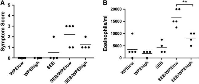
Results: SEB administered with antigen resulted in immune responses to the antigen. Responses were highly T(H)2 polarized, and oral challenge with antigen triggered anaphylaxis and local and systemic mast cell degranulation. SEB-driven sensitization induced eosinophilia in the blood and intestinal tissues not observed with cholera toxin sensitization. SEB impaired tolerance specifically by impairing expression of TGF-beta and regulatory T cells, and tolerance was restored with high-dose antigen.

Conclusions: We demonstrate a new model of food allergy to oral antigen in common laboratory strains of mice that recapitulates many features of clinical food allergy that are not seen in other models. We demonstrate that SEB impairs oral tolerance and permits allergic responses.


Figures

FIG 1
FIG 1
Antigen-driven anaphylaxis and mast cell activation in SEB/OV A-sensitized mice. After sensitization, mice received a bolus dose of 5 mg of OVA by mouth. A, Symptom scores elicited in CT-driven or SEB-driven sensitization were determined. B, SEB/OVA-sensitized mice responded to OVA but not to an irrelevant antigen, BSA. C, Plasma histamine levels were determined 60 minutes after antigen challenge. D, Numbers and granulation status of mast cells in ear skin were determined by means of histologic analysis. E, A representative section showing pinacyanol erythrosinate staining on jejunum tissue from an SEB/OVA mouse after challenge is shown. Mast cells are stained with a deep blue/purple color. F, Hematoxylin and eosin staining of jejunum , illustrating vasodilation. G, Blood pressure was measured in OVA/SEB-treated mice to determine systemic responses immediately after antigen challenge with a Coda 1 noninvasive blood pressure system . In Fig 1, C and D, data represent the means 6 SEMs (n 5 6-12). *P < .05 and **P < .01, Student t test.
FIG 2
FIG 2
SEB-driven sensitization promotes increased eosinophilia, whereas CT does not. Peripheral blood eosinophil numbers were determined in treated mice. A, Peripheral blood eosinophils were increased in SEB/OVA-treated mice but not in CT/OVA-treated mice. B, Eosinophilia correlated with the amount of SEB administered during sensitization. C, Representative history of tissue stained with hematoxylin and eosin demonstrating a robust eosinophilic infiltration into the jejunum tissues. Black arrows indicate eosinophils. *P < .05 and **P < .01, Student t test (n 5 6-12).
FIG 3
FIG 3
A TH2-skewed antigen-dependent cytokine profile develops after SEB-driven sensitization. Splenocytes from mice previously sensitized with SEB alone, OVA alone, or SEB/OVA were stimulated for 48 hours in the presence of 100 mg of OVA or with plate-bound anti-mouse CD3 (1 mg/mL). A, Cytokine levels in the supernatants were determined by using the cytometric bead assay. Gene expression levels were determined for TH/TH2 (B) and Treg cell–associated (C) mediators. *P < .05 and **P < .01, Student t test (n 5 5-6). NS, No significant difference.
FIG 4
FIG 4
SEB promotes allergic responses to peanut at a low dose but not at a high dose. Responses to oral administration of 100 mg of WPE (WPElow) or 1 mg of WPE (WPEhigh) 6 10 mg of SEB were determined. Symptom scoring in response to challenge with 5 mg of WPE (A), peripheral blood eosinophilia (B), and WPE-specific IgE levels (C) was determined. Finally, in vitro cytokines on stimulation of splenocytes with 1 mg of WPE or anti-mouse CD3 (1 mg/mL) for 48 hours were studied. IL-4 (D), IL-5 (E), and IFN-g (F) levels are shown. *P < .05 and **P < .01. Student t test (n 5 5 per group).
FIG E1
FIG E1
T-cell receptor repertoire use after oral SEB exposure. The percentage of each Vb subset of T cells was determined by using flow cytometry on spleen cells. Data represent the average percentage of Vb subsets for the entire CD41 T-cell population. **P < .01, Student t test (n 5 5 per group).
FIG E2
FIG E2
Core temperatureresponses after antigen challenge. Rectal temperature was measured in WPE/SEB-treated mice to determine systemic responses immediately after antigen challenge. Rectal temperatures were measured concurrently in 7 animals by using an automated PhysioTemp mouse rectal temperature system (PhysioTemp Instruments, Inc, Clifton, NJ) and shown as a change in baseline for each individual mouse, and these were not significantly different (36.78C 6 0.48C for control animals and 36.28C 6 0.48C for sensitized mice).
FIG E3
FIG E3
SEB promotes allergic responses to peanut in C57BL/6J mice. Responses to oral administration of 100 mg of WPE (WPElow) or 1 mg of WPE (WPEhigh) 6 10 mg of SEB were determined. Symptom scoring in response to challenge with 5 mg of WPE (A) and peripheral blood eosinophilia (B) are shown. **P < .01, Student t test.

References

    1. Lee SY, Huang CK, Zhang TF, Schofield BH, Burks AW, Bannon GA, et al. Oral administration of IL-12 suppresses anaphylactic reactions in a murine model of peanut hypersensitivity. Clin Immunol. 2001;101:220–8. - PubMed
    1. Li XM, Zhang TF, Huang CK, Srivastava K, Teper AA, Zhang L, et al. Food Allergy Herbal Formula-1 (FAHF-1) blocks peanut-induced anaphylaxis in a murine model. J Allergy Clin Immunol. 2001;108:639–46. - PubMed
    1. Srivastava KD, Kattan JD, Zou ZM, Li JH, Zhang L, Wallenstein S, et al. The Chinese herbal medicine formula FAHF-2 completely blocks anaphylactic reactions in a murine model of peanut allergy. J Allergy Clin Immunol. 2005;115:171–8. - PubMed
    1. van Wijk F, Hartgring S, Koppelman SJ, Pieters R, Knippels LM. Mixed antibody and T cell responses to peanut and the peanut allergens Ara h 1, Ara h 2, Ara h 3 and Ara h 6 in an oral sensitization model. Clin Exp Allergy. 2004;34:1422–8. - PubMed
    1. van Wijk F, Hoeks S, Nierkens S, Koppelman SJ, van Kooten P, Boon L, et al. CTLA-4 signaling regulates the intensity of hypersensitivity responses to food antigens, but is not decisive in the induction of sensitization. J Immunol. 2005;174:174–9. - PubMed

Publication types

MeSH terms