Review
. 2008:73:333-50.
doi: 10.1101/sqb.2008.73.032. Epub 2008 Nov 6.

Building epithelial tissues from skin stem cells

Affiliations
Review

Building epithelial tissues from skin stem cells

E Fuchs et al. Cold Spring Harb Symp Quant Biol. 2008.

Abstract

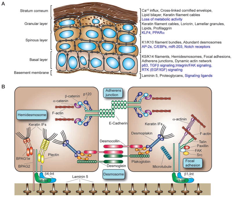
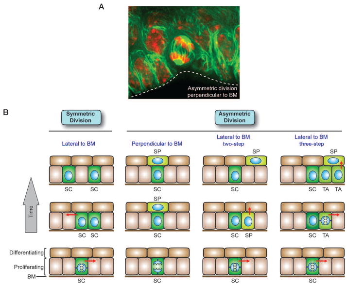
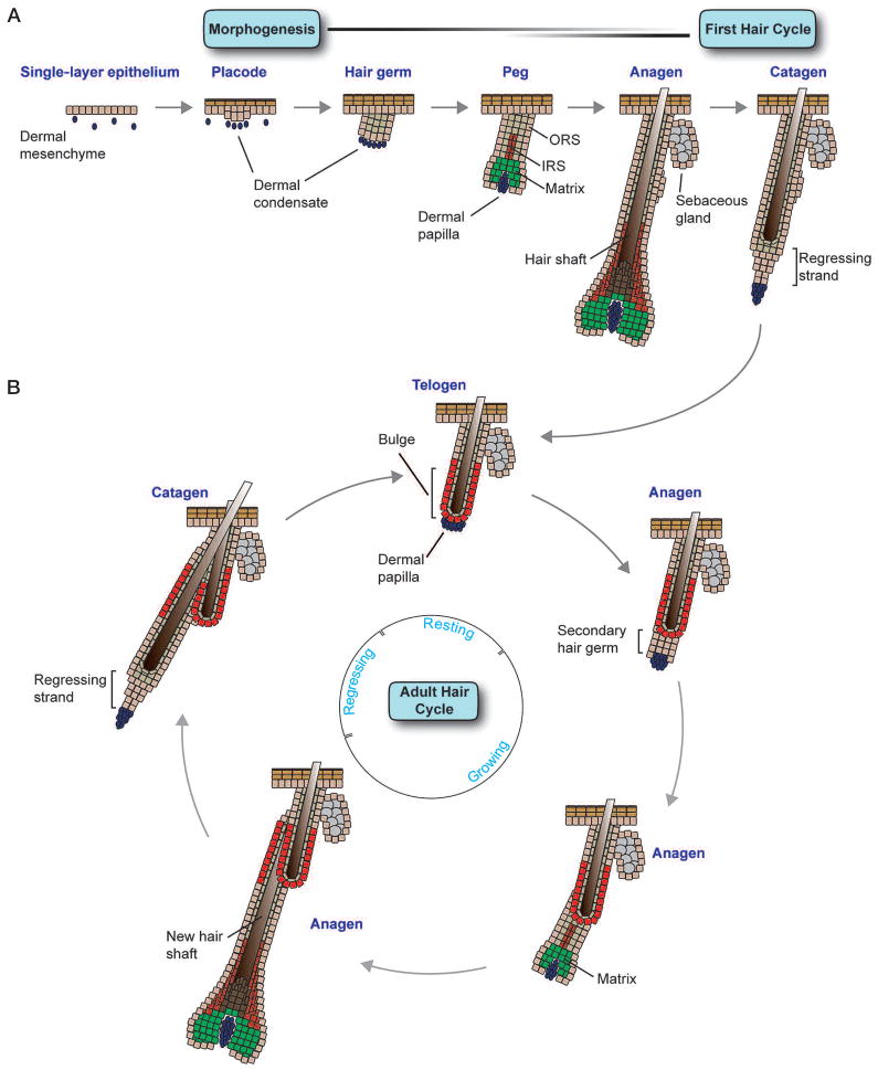
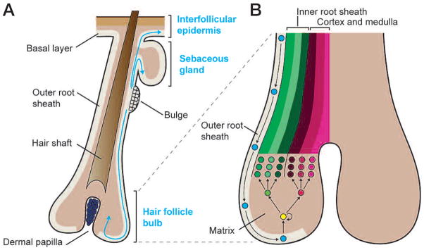
The skin epidermis and its appendages provide a protective barrier that guards against loss of fluids, physical trauma, and invasion by harmful microbes. To perform these functions while confronting the harsh environs of the outside world, our body surface undergoes constant rejuvenation through homeostasis. In addition, it must be primed to repair wounds in response to injury. The adult skin maintains epidermal homeostasis, hair regeneration, and wound repair through the use of its stem cells. What are the properties of skin stem cells, when do they become established during embryogenesis, and how are they able to build tissues with such remarkably distinct architectures? How do stem cells maintain tissue homeostasis and repair wounds and how do they regulate the delicate balance between proliferation and differentiation? What is the relationship between skin cancer and mutations that perturbs the regulation of stem cells? In the past 5 years, the field of skin stem cells has bloomed as we and others have been able to purify and dissect the molecular properties of these tiny reservoirs of goliath potential. We report here progress on these fronts, with emphasis on our laboratory's contributions to the fascinating world of skin stem cells.


Figures

Figure 1
Figure 1
The cellular architecture of the epidermis and major components of the epidermal cytoskeleton. (A) Program of epidermal differentiation illustrating the BM at the base, the proliferative basal layer, and the three differentiation stages: spinous layer, granular layer, and outermost stratum corneum. Shown at the right are key molecular markers described in the text. (Black text) Structural and cytoskeletal components present in each layer; (purple text) regulatory mechanisms and signaling activity necessary for proper function of each layer. (B) Key components of the cytoskeleton in basal layer keratinocytes. Keratin IFs connect to the BM via hemidesmosomes, whose core components include homodimers of the plakin proteins plectin and BPAG1e, the transmembrane collagen BPAG2, and a heterodimer composed of α6 and β4 integrin. Intercellularly, the keratin network is also linked at adjacent cells by desmosomes that are composed of the desmosomal cadherins desmocollin and desmoglein, the linker protein plakoglobin, and a homodimer of the plakin protein desmoplakin. Analogously, the actin cytoskeleton attaches to the underlying BM through focal adhesions that are composed of an α3β1 integrin heterodimer core linked to actin filaments (F-actin) by talin and α-actinin. Paxillin, FAK, and Src are also associated with focal adhesions and help to orchestrate signaling events regulating cell adhesion and motility. Microtubules can also contact focal adhesions, helping to regulate their turnover. Between adjacent cells, the actin cytoskeleton is linked through adherens junctions that associate through homotypic interactions between E-cadherin molecules. Heterodimers composed of β-catenin and α-catenin regulate the link between E-cadherin and the actin cytoskeleton, whereas p120-catenin stabilizes E-cadherin interactions by binding to E-cadherin’s cytoplasmic domain.
Figure 2
Figure 2
Roles for symmetric and asymmetric cell division in epidermal development and homeostasis. (A) Immunofluorescence image of a cell in the basal layer of an E15.5 embryonic tongue undergoing asymmetric division perpendicular to the BM (dotted white line). The microtubule network is marked by green fluorescent protein (GFP), and DNA is marked by red propidium iodide. (B) Self-renewing stem cells (SC) exist in the basal layer of the epidermis. Symmetrical divisions lateral to the BM produce two stem cells, a process that can serve to refill vacancies in the basal layer or increase the area of the epidermis during development. Asymmetric divisions can occur both laterally and perpendicular to the BM. In the two-step asymmetric division model, a stem cell divides asymmetrically to preferentially partition proliferation-associated factors into the stem cell daughter and provide differentiation-inducing components to the other daughter, fated to become a spinous (SP) cell. If the spindle orientation was perpendicular to the BM, the division could result in direct positioning of the SP daughter away from the BM, whereas lateral spindle orientation relative to the BM would then necessitate subsequent delamination of the committed SP daughter. In the three-step asymmetric division model, a transit-amplifyng (TA) intermediate arises, which has been postulated to divide three to four times before delaminating (arrows) and entering into a terminal differentiation program. Once the spinous cells have separated from the BM, they enter a program of terminal differentiation as they move outward and are eventually sloughed from the skin surface (see Fig. 1). Differentiating cells are continually replaced by a flux of inner cells committing to terminally differentiate and move outward. Immunofluorescence image courtesy of Terry Lechler (when in the Fuchs lab).
Figure 3
Figure 3
Embryonic hair follicle morphogenesis and the adult hair cycle. (A) The process of follicle morphogenesis occurs in several overlapping waves that begin at E14.5 when small invaginations termed placodes appear in the basal layer of the epidermis, accompanied by aggregations of dermal cells termed dermal condensates. Cells of the placode proliferate and grow downward into the dermis to form hair germs (~E16.5–E17.5). Next, during the peg stage (~E18.5–P0), transit-amplifying matrix cells appear at the base of the follicle and encapsulate the dermal papilla (DP). The upper portion of the follicle also becomes separated into the outer root sheath (ORS) and inner root sheath (IRS) at this time. Soon thereafter (~P1–P3), fully differentiated hair shafts and sebaceous glands (SGs) appear, and follicle morphogenesis reaches completion at about P9. At this time, follicles reach their maximum length, marking entrance into the anagen phase of the first hair cycle. After production of the first hair coat, follicles transition to the catagen phase of the hair cycle (~P16–P19), where the matrix undergoes apoptosis and degenerates into a regressing epithelial strand that draws the DP upward to rest just below the base of the first “club” hair, which is surrounded by the newly formed adult bulge niche. (B) Direct contact between the DP and adult bulge cells indicates the onset of telogen (~P20). Soon thereafter, at about P21, bulge stem cells are activated and a secondary hair germ grows downward from the base of the bulge niche, signaling entry into the anagen phase of the next hair cycle. The emergence of a new hair follicle from the side of the original club hair lends the stem cell niche its characteristic “bulge” morphology and divides the CD34-positive stem cells into two populations based on high or low integrin expression levels and adherence to the BM. In a process that has many similarities to that of initial hair follicle morphogenesis, the secondary germ expands and gives rise to a new matrix that begins to produce the differentiated lineages of the IRS and hair shaft. This new hair shaft then exits from the same channel as the existing club hair. After several weeks of growth, anagen ceases and follicles enter catagen, again drawing the DP upward to rest below the bulge stem cell niche. After a variable period of time, bulge stem cells are again activated to initiate a new hair growth cycle. For a comprehensive analysis of the details and classification of hair follicle morphogenesis and adult hair cycling, see Muller-Rover et al. (2001), Stenn and Paus (2001), and Schmidt-Ullrich and Paus (2005).
Figure 4
Figure 4
Multipotency of bulge epithelial stem cells and lineage decisions in the hair follicle. (A) Epithelial stem cells reside in a specialized niche in the upper ORS of each hair follicle and can give rise to all three epithelial lineages of the skin. During normal homeostasis, bulge stem cells are periodically activated to form a new hair follicle. During the hair follicle growth period (anagen), bulge cells migrate down the lower ORS toward the matrix, which is a specialized population of highly proliferative transit-amplifying cells responsible for producing a new hair. Bulge stem cells can also migrate to and differentiate along an SG lineage when sebaceous progenitors are absent or impaired. In a wound environment, bulge stem cells can also migrate upward and out of the hair follicle to contribute to regeneration of the interfollicular epidermis. (B) As bulge progeny migrate down the ORS, they subsequently enter the matrix. Matrix cells then detach from the BM and differentiate along one of six hair lineages, three of which comprise the IRS and three the cortex and medulla of the hair shaft. Intimate contact with the dermal papilla is essential for maintaining the high proliferative capacity of the matrix and driving lineage decisions.
Figure 5
Figure 5
Regulation of stem cell identity and activity in the adult bulge stem cell niche. (Top) In telogen-phase hair follicles, stem cells residing in the bulge are quiescent and express the markers CD34, Sox9, Tcf3, Nfatc1, and Lhx2. High levels of BMP signaling maintain stem cells in a quiescent state, whereas low levels of Wnt signaling may help to maintain stem cell identity but are insufficient to drive SC activation. (Bottom) In early anagen-phase hair follicles, stem cells in the bulge proliferate and give rise to a secondary hair germ that loses most stem cell markers but still retains Sox9 and Lhx2 expression. In contrast to the bulge, BMP signaling is down-regulated and Wnt signaling is up-regulated in the germ, allowing cells to proliferate rapidly in order to produce a new hair follicle.
Figure 6
Figure 6
Embryonic specification of the hair follicle stem cell population. (A) Sox9, a gene essential for specifying the hair follicle stem cell population, is expressed in suprabasal cells of the placode at the first stage of hair follicle morphogenesis. As development proceeds, Sox9 remains in the upper portion of the ORS, marking the early follicle stem cell population, as well as in their progeny that migrate down the ORS toward the matrix. In adult follicles, Sox9 remains expressed in the bulge stem cell niche. (B) Genetic marking studies of Sox9-expressing cells show that hair follicles are initially composed of two cell populations, Sox9-expressing early stem cells and transient Sox9-negative cells that give rise to the initial matrix. As follicle morphogenesis proceeds, the progeny of Sox9-expressing cells move down the ORS and completely replace the initial matrix population. Additionally, progeny of Sox9-positive cells give rise to the SG lineage and can help repair the interfollicular epidermis in a wound environment. By the time hair follicle morphogenesis has finished, hair follicles are entirely derived from Sox9-expressing cells. (C) Markers and functional characteristics of the hair follicle stem cell population are acquired in a stepwise manner. The transcription factors Lhx2 and Sox9 are expressed at the first placode stage of morphogenesis, whereas Tcf3 and Nfatc1 appear later at the hair germ stage. Although all four of these genes mark both early and adult stem cells, CD34 is only up-regulated in adult follicle stem cells. Morphologically, the location of early stem cells in the follicle can be inferred by a thickening in the ORS that appears during late stages of morphogenesis and becomes much more pronounced when the adult bulge stem cell niche forms during the first telogen. Although all early stem cells divide at least several times during follicle morphogenesis, they gradually increase their slow-cycling character as the rate of hair follicle growth decreases. Adult stem cells in resting telogen hair follicles are highly quiescent, but they undergo periods of reduced quiescence in the growing anagen phase of the hair cycle.

Similar articles

Cited by

References

    1. Alonso L, Fuchs E. The hair cycle. J Cell Sci. 2006;119:391–393. - PubMed
    1. Andl T, Reddy ST, Gaddapara T, Millar SE. WNT signals are required for the initiation of hair follicle development. Dev Cell. 2002;2:643–653. - PubMed
    1. Andl T, Ahn K, Kairo A, Chu EY, Wine-Lee L, Reddy ST, Croft NJ, Cebra-Thomas JA, Metzger D, Chambon P, et al. Epithelial Bmpr1a regulates differentiation and proliferation in postnatal hair follicles and is essential for tooth development. Development. 2004;131:2257–2268. - PubMed
    1. Arnold I, Watt FM. c-Myc activation in transgenic mouse epidermis results in mobilization of stem cells and differentiation of their progeny. Curr Biol. 2001;11:558–568. - PubMed
    1. Balzac F, Avolio M, Degani S, Kaverina I, Torti M, Silengo L, Small JV, Retta SF. E-cadherin endocytosis regulates the activity of Rap1: A traffic light GTPase at the crossroads between cadherin and integrin function. J Cell Sci. 2005;118:4765–4783. - PubMed

Publication types

MeSH terms