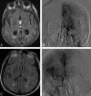
Angiography reveals that fluid-attenuated inversion recovery vascular hyperintensities are due to slow flow, not thrombus
- PMID: 19022866
- PMCID: PMC2729168
- DOI: 10.3174/ajnr.A1388
Angiography reveals that fluid-attenuated inversion recovery vascular hyperintensities are due to slow flow, not thrombus
Abstract
Background and purpose: Fluid-attenuated inversion recovery (FLAIR) vascular hyperintensities (FVH) are commonly encountered on MR imaging studies performed shortly after the onset of acute ischemic stroke. Prior reports have speculated regarding the pathogenesis of this finding, yet definitive correlative angiographic studies have not been performed. We studied the pathophysiologic and hemodynamic correlates of FVH on conventional angiography and concurrent MR imaging sequences.
Materials and methods: Retrospective review of FLAIR and gradient-refocused echo MR imaging sequences acquired immediately before conventional angiography for acute stroke was conducted in a blinded fashion. The presence, location, and morphology of FVH were noted and correlated with markers of thrombotic occlusion and collateral flow on angiography. Angiographic collaterals were graded on a 5-point scale incorporating extent and hemodynamic aspects.
Results: A prospective ischemic stroke registry of 632 patients was searched to identify 74 patients (mean age, 63.4 +/- 20 years; 48% women) having undergone FLAIR sequences immediately before angiography. Median time from FLAIR to angiography was 2.9 hours (interquartile range, 1.1-4.7 hours). FVH were present in 53/74 (72%) of all acute stroke cases with subsequent angiography. FVH distal to an arterial occlusion were associated with a high grade of leptomeningeal collateral blood flow.
Conclusions: FVH are observed in areas of blood flow proximal and distal to stenosis or occlusion and are noted with more extensive collateral circulation.
Figures


References
-
- Kamran S, Bates V, Bakshi R, et al. Significance of hyperintense vessels on FLAIR MRI in acute stroke. Neurology 2000;55:265–69 - PubMed
-
- Tsushima Y, Endo K. Significance of hyperintense vessels on FLAIR MRI in acute stroke. Neurology 2001;56:1248–49 - PubMed
-
- Iancu-Gontard D, Oppenheim C, Touze E, et al. Evaluation of hyperintense vessels on FLAIR MRI for the diagnosis of multiple intracerebral arterial stenoses. Stroke 2003;34:1886–91 - PubMed
Publication types
MeSH terms
Grants and funding
LinkOut - more resources
Full Text Sources
