. 2009 Apr;150(4):1841-9.
doi: 10.1210/en.2008-1023. Epub 2008 Nov 20.

Prolactin activates mitogen-activated protein kinase signaling and corticotropin releasing hormone transcription in rat hypothalamic neurons

Affiliations

Prolactin activates mitogen-activated protein kinase signaling and corticotropin releasing hormone transcription in rat hypothalamic neurons

Annegret Blume et al. Endocrinology. 2009 Apr.

Abstract

Prolactin (PRL) modulates maternal behavior and mediates hypothalamic pituitary adrenal axis inhibition during lactation via PRL receptors in the brain. To identify mechanisms mediating these effects, we examined the effects of PRL on signaling and CRH transcription in hypothalamic neurons in vivo and in vitro. Western blot of hypothalamic proteins from rats receiving intracerebroventricular PRL injection revealed increases in phosphorylation of the MAPK and ERK. Double-staining immunohistochemistry demonstrated phosphorylated ERK localization in parvocellular CRH neurons as well as magnocellular vasopressin and oxytocin neurons of the hypothalamic paraventricular (PVN) and supraoptic nuclei. PRL also induced ERK phosphorylation in vitro in the hypothalamic cell line, 4B, which expresses PRL receptors, and in primary hypothalamic neuronal cultures. Using reporter gene assays in 4B cells, or quantitative RT-PCR for primary transcript in hypothalamic cell cultures, PRL potentiated forskolin-stimulated CRH transcription through activation of the ERK/MAPK pathway. The effect of PRL in hypothalamic cell cultures was unaffected by tetrodotoxin, suggesting a direct effect on CRH neurons. The data show that PRL activates the ERK/MAPK pathway and facilitates CRH transcription in CRH neurons, suggesting that the inhibitory effect of PRL on hypothalamo-pituitary-adrenal axis activity reported in vivo is indirect and probably mediated through modulation of afferent pathways to the PVN. In addition, the prominent stimulatory action of PRL on the ERK/MAPK pathway in the hypothalamic PVN and supraoptic nucleus is likely to mediate neuroplasticity of the neuroendocrine system during lactation.


Figures

Figure 1
Figure 1
Activation of the ERK1/2 MAPK pathway by PRL in the rat hypothalamus. PRL (1 μg) or vehicle (Veh; n = 5 animals/group) were injected into the lateral brain ventricle of conscious virgin female rats. Five, 10, or 30 min later, the hypothalami were microdissected and the cytoplasmatic and nuclear proteins extracted. Proteins were analyzed by Western blot using phospho-specific primary antibodies (pMEK1/2, pERK1/2, control MEK1/2, ERK1/2, 1:1000), followed by signal enhancement with a peroxidase-coupled secondary antibody (antirabbit, 1:1000) and visualization with enhanced chemiluminescence. A, Cytoplasmatic pMEK. B, Cytoplasmatic pERK1/2. C, Nuclear translocation of pERK, *, P < 0.05, **, P < 0.01 vs. Veh; inserts, representative Western blots. D, Immunohistochemical staining for pERK in rats 10 min after icv injection of vehicle (Veh) or 1 μg PRL in the hypothalamic PVN and SON regions. Staining was absent in sections in which the primary antibody was omitted (not shown). 3V, Third ventricle; OC, optic chiasm.
Figure 2
Figure 2
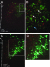
Colocalization of CRH and pERK in the hypothalamic PVN. Conscious virgin female rats (three per group) received a PRL injection (1 μg, icv) and were killed 10 min later. For visualization of colocalization of pERK with CRH, incubation with two primary antibodies (pERK mouse monoclonal antibody E10, 1:100; CRH rabbit polyclonal antibody, 1:1000) was followed by a combination of secondary antibodies [goat-antimouse IgG, Alexa Fluor 488 (green, pERK); goat-antirabbit IgG, Alexa Fluor 546 (red, CRH)]. Whereas CRH staining (A) is found in the cytosol, pERK staining (B) was present predominantly in the nucleus but also in the cytosol. The overlap of the CRH and pERK staining is shown in C.
Figure 3
Figure 3
Colocalization of PRL-induced pERK1/2 with VP and OT in the hypothalamic PVN. Conscious rats were treated with PRL (1 μg, icv) for 10 min (n = 3 animals/group). For colocalization with VP and OT, pERK was visualized on 4-μm brain slices using two primary antibodies (pERK1/2 rabbit monoclonal antibody, 1:500; VP mouse monoclonal antibody, 1:500 or OT mouse monoclonal antibody, 1:1000) followed by a combination of secondary, fluorescence coupled-antibodies [goat-antirabbit IgG, Alexa Fluor 546 (red; pERK); goat-antimouse-IgG, Alexa Fluor 488 (green; VP and OT)]. A, pERK/VP. B, Detail of A. C, pERK/OT. D, Detail of D. 3V, Third ventricle; white arrows, double-positive neurons.
Figure 4
Figure 4
Colocalization of PRL-induced pERK1/2 with VP (upper panel) and OT (lower panel) in the hypothalamic SON. Conscious rats were treated with PRL (1 μg, icv) for 10 min (n = 3 animals/group). For colocalization with VP and OT, pERK was visualized on 4-μm brain slices using two primary antibodies (pERK1/2 rabbit monoclonal antibody, 1:500; VP mouse monoclonal antibody, 1:500 or OT mouse monoclonal antibody, 1:1000) followed by a combination of secondary, fluorescence coupled-antibodies (for details see legend to Fig. 3) Left, pERK; middle: VP (upper) and OT (lower); right: pERK+VP (upper) and pERK+OT (lower). OC, Optic chiasm; white arrows, double-labeled cells.
Figure 5
Figure 5
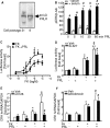
Effects of PRL on ERK1/2 phosphorylation and CRH transcription in hypothalamic cells. A, Western blot for the PRL receptor in protein extracts from the hypothalamic cell line 4B on passages 21 and 8. A specific 40-kDa band corresponding to the molecular mass of the short form of PRL receptor was seen at both passages. B, Time course of the effect of PRL (1 μg/ml) on pERK1/2 levels in the presence or absence of serum in 4B cells measured by Western blot using a phospho-specific antibody. Bars represent the mean and se of the values in five experiments. *, P < 0.05, **, P < 0.01 vs. respective time 0; #, P < 0.001 vs. respective serum free. C, Dose-response of the effect of forskolin (FK) with or without prolactin on CRH promoter activity in 4B cells transiently transfected with a CRH promoter-driven luciferase construct. Eighteen hours after transfection, cells were incubated for 6 h with FK (0.05–10 μm) in the absence or presence of PRL (1 μg/ml). Data points are the mean and se of the results in five experiments. *, P < 0.05 vs. respective dose of FK alone. D, Eighteen hours after transfection, cells were preincubated for 30 min with the MEK inhibitor SL327 (10 μm), followed by incubation with Veh, FK (0.3 μm), PRL (1 μg/ml), or a combination of FK (0.3 μm) and PRL (1 μg/ml) in the presence or absence of SL327 (10 μm) for 6 h (n = 5 experiments per data point). *, P < 0.05 vs. basal (Veh, Veh+SL) or PRL with or without SL327; **, P < 0.001 vs. basal or PRL with or without SL327; #, P < 0.05 vs. FK+PRL; §, P < 0.01 vs. FK with or without SL327. E, Effect of FK with or without PRL on CRH hnRNA levels in the presence or absence of the MEK inhibitor, U0126 in primary hypothalamic cultures. Cells were pretreated with either vehicle or the MEK inhibitor, U0126, before addition of vehicle (Veh), forskolin (FK) (0.3 μm), PRL (1 μg/ml), or their combination for 45 min, measured by intronic quantitative RT-PCR. Bars represent the mean and se of the values obtained in five experiments. *, P < 0.05 vs. Veh; **, P < 0.01 vs. basal (Veh, Veh+U0126) or PRL with or without U0126; #, P < 0.05 vs. FK+PRL; §, P < 0.05 vs. FK with or without U0126 or PRL with or without U0126; &, P < 0.05 vs. PRL. F, Effect of the sodium channel blocker, tetrodotoxin, on the effect of PRL on CRH hnRNA in hypothalamic neuronal cultures. Cells were pretreated with either Veh or tetrodotoxin before addition of FK (0.3 μm), PRL (1 μg/ml), or FK+ PRL and incubation for an additional 45 min. Tetrodotoxin had no effect on the FK-induced increase in CRH hnRNA in the absence or in the presence of PRL. *, P < 0.01 vs. respective basal; P < 0.05 vs. FK+PRL; #, P < 0.01 vs. respective FK alone.

Similar articles

Cited by

References

    1. Mangurian LP, Walsh RJ, Posner BI 1992 Prolactin enhancement of its own uptake at the choroid plexus. Endocrinology 131:698–702 - PubMed
    1. Walsh R, Slaby F, Posner B 1987 A receptor-mediated mechanism for the transport of prolactin from blood to cerebrospinal fluid. Endocrinology 120:1846–1850 - PubMed
    1. Ben-Jonathan N, Mershon JL, Allen DL, Steinmetz RW 1996 Extrapituitary prolactin: distribution, regulation, functions, and clinical aspects. Endocr Rev 17:639–669 - PubMed
    1. Clapp C, Torner L, Gutierrez-Ospina G, Alcantara E, Lopez-Gomez F, Nagano M, Kellly P, Mejia S, Ma M, Martinez de la Escalera G 1994 The prolactin gene is expressed in the hypothalamic-neurohypophyseal system and the protein is processed into a 14-kDa fragment with activity like 16-kDa prolactin. Proc Natl Acad Sci USA 91:10384–10388 - PMC - PubMed
    1. Emanuele N, Jurgens J, Halloran M, Tentler J, Lawrence A, Kelley M 1992 The rat prolactin gene is expressed in brain tissue: detection of normal and alternatively spliced prolactin messenger RNA. Mol Endocrinol 6:35–42 - PubMed

Publication types

MeSH terms

Substances