. 2008 Dec 2;105(48):18758-63.
doi: 10.1073/pnas.0805411105. Epub 2008 Nov 20.

Perturbation of transforming growth factor (TGF)-beta1 association with latent TGF-beta binding protein yields inflammation and tumors

Affiliations

Perturbation of transforming growth factor (TGF)-beta1 association with latent TGF-beta binding protein yields inflammation and tumors

Keiji Yoshinaga et al. Proc Natl Acad Sci U S A. .

Abstract

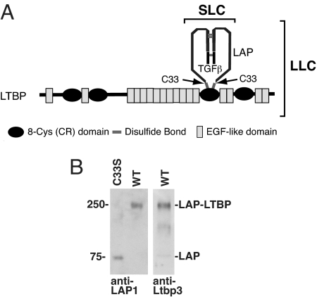
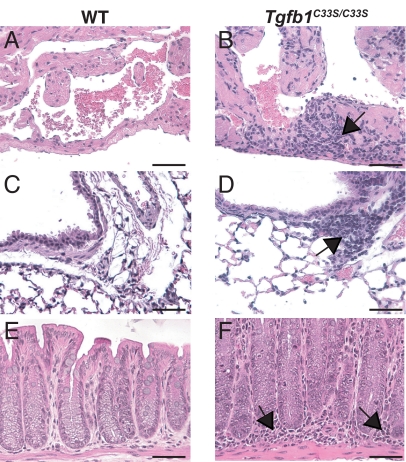
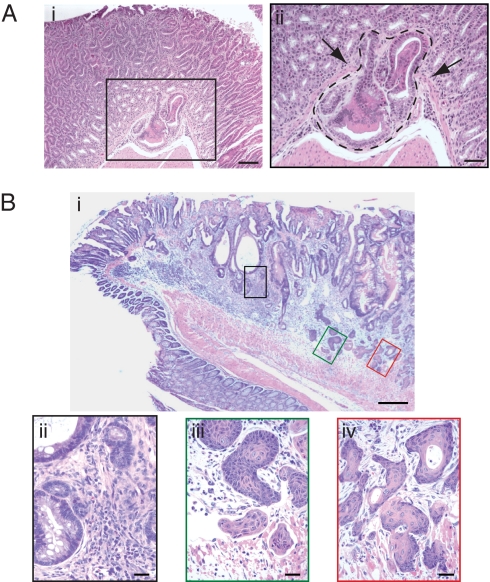
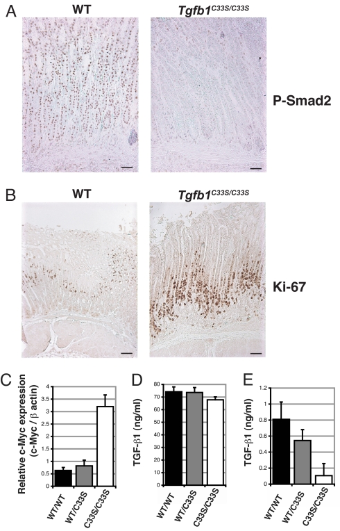
Transforming growth factor-beta (TGF-beta) activity is controlled at many levels including the conversion of the latent secreted form to its active state. TGF-beta is often released as part of an inactive tripartite complex consisting of TGF-beta, the TGF-beta propeptide, and a molecule of latent TGF-beta binding protein (LTBP). The interaction of TGF-beta and its cleaved propeptide renders the growth factor latent, and the liberation of TGF-beta from this state is crucial for signaling. To examine the contribution of LTBP to TGF-beta function, we generated mice in which the cysteines that link the propeptide to LTBP were mutated to serines, thereby blocking covalent association. Tgfb1(C33S/C33S) mice had multiorgan inflammation, lack of skin Langerhans cells (LC), and a shortened lifespan, consistent with decreased TGF-beta1 levels. However, the inflammatory response and decreased lifespan were not as severe as observed with Tgfb1(-/-) animals. Tgfb1(C33S/C33S) mice exhibited decreased levels of active TGF-beta1, decreased TGF-beta signaling, and tumors of the stomach, rectum, and anus. These data suggest that the association of LTBP with the latent TGF-beta complex is important for proper TGF-beta1 function and that Tgfb1(C33S/C33S) mice are hypomorphs for active TGF-beta1. Moreover, although mechanisms exist to activate latent TGF-beta1 in the absence of LTBP, these mechanisms are not as efficient as those that use the latent complex containing LTBP.


Conflict of interest statement

The authors declare no conflict of interest.

Figures

Fig. 1.
Fig. 1.
TGF-β1 latent complexes from Tgfb1+/+ and Tgfb1C33S/C33S mice. (A) Structure of latent TGF-β LLC. The LLC of TGF-β1 consists of the mature cytokine dimer, the propeptide dimer (LAP), and LTBP. The LAP residues that bind to LTBP are cysteines 33. Changing this residue to serine prevents formation of the TGF-β1 LLC. (B) LAP associations in Tgfb1+/+ and Tgfb1C33S/C33S lung cell culture media as revealed after SDS/PAGE and immunoblotting. Immunoreactive material from Tgfb1C33S/C33S cells is detected only at the position of the LAP dimer (75 kDa). (Left) There is no band at the position of LAP-LTBP. LAP immunoreactive material is present at the position corresponding to 250 kDa, the position of LAP plus LTBP. (Center) Western blot of Tgfb1C33S/C33S cell conditioned medium with an antibody to LTBP-3. (Right) The positive reaction indicates that the LLC is composed of LAP and LTBP-3. Blotting with antibodies to LTBP-1 revealed no LLC indicating the absence or undetectable levels of this species in either sample. Antibody to LTBP-4 was not tested.
Fig. 2.
Fig. 2.
Inflammation in Tgfb1+/+ and Tgfb1C33S/C33S tissues. Samples were taken from WT and Tgfb1C33S/C33S fixed, sectioned, and stained. (A, C, and E) Tgfb1+/+ tissues. (B, D, and E) Tgfb1C33S/C33S tissues. (A and B) Heart. There is no inflammation in the Tgfb1+/+ heart, but inflammatory cells (arrows) are present in the mutant heart. (C and D) Lung. Although the numbers of inflammatory cells in the Tgfb1+/+ lung are low, in the mutant tissue significant numbers of inflammatory cells surround certain blood vessels (arrows). (E and F) Terminal colon/rectum. The tissue from the Tgfb1+/+ mouse displays few inflammatory cells. However, the tissue from the Tgfb1C33S/C33S animal had large numbers of inflammatory cells in the lamina propria. The heart and lung tissues were from 4-week-old animals and the terminal colon/rectal tissue from 12-week-old mice. (Scale bar: 50 μM.)
Fig. 3.
Fig. 3.
Tumors in Tgfb1C33S/C33S mice. (Ai) Section from the glandular stomach mucosa illustrating an early gastric adenocarcinoma. (ii) Higher magnification of the enclosed box in Ai. Arrows point to invasive dysplastic epithelial cells that have breached the basement membrane and muscularis mucosa. (B) (i) Section near the rectal–anal junction illustrating an adenosquamous carcinoma. (ii–iv) Higher magnification of regions comprising the boxed areas shown in B. (Biii) Tumor with adenomatous histology. (iii–iv) Tumor with squamous cell carcinoma histology. [Scale bars: 100 μM (Ai); 50 μM (Aii); 200 μM (Bi); 50 μM (Bii); and 20 μM (B iii and iv).]
Fig. 4.
Fig. 4.
TGF-β signaling and mitosis in tissues from Tgfb1+/+ and Tgfb1C33S/C33S mice. (A) P-Smad2 staining of stomach from Tgfb1+/+ and Tgfb1C33S/C33S of 11-week-old mice. The more intense P-Smad2 nuclear staining in the Tgfb1+/+ vs. the Tgfb1C33S/C33S sample indicates enhanced TGF-β activity. (B) Staining of stomach tissue from Tgfb1+/+ and Tgfb1C33S/C33S 21-week-old mice with antibody to Ki67, a marker of mitosis. The number of positive Ki67 cells is higher in the mutant sample compared to the Tgfb1+/+ sample, consistent with decreased TGF-β1 in the tissue, as TGF-β1 is a suppressor of growth of most epithelial cells. (C) Q-RT-PCR of stomach epithelium for Myc from 12-week-old mice. Tgfb1C33S/C33S tissue had higher expression of Myc than did control tissue consistent with decreased TGF-β1 in the mutant tissue. (D and E) TGF-β1 in sera from Tgfb1+/+ and Tgfb1C33S/C33S mice. TGF-β1 in sera from Tgfb1+/+, Tgfb1+/C33S, and Tgfb1C33S/C33S mice was measured by an ELISA specific for active TGF-β1. (D) Total TGF-β1 in sera after acidification. Acidification releases all active TGF-β from its latent complex. The total TGF-β1 from the three genotypes was equivalent. (E) Active TGF-β1 in sera. TGF-β1 was measured directly in sera without acidification. The amount of active TGF-β1 in the Tgfb1+/+ and Tgfb1+/C33S samples is significantly higher than that found in samples from Tgfb1C33S/C33S mice. Samples from 10–12 animals of each genotype were assayed for TGF-β. Assays on each serum sample were done in triplicate. Values are presented as SEM.

References

    1. Annes JP, Munger JS, Rifkin DB. Making sense of latent TGFbeta activation. J Cell Sci. 2003;116:217–224. - PubMed
    1. Derynck R, Miyazono K. The TGF-â Family. Cold Spring Harbor, NY: Cold Spring Harbor Lab Press; 2008. - PMC - PubMed
    1. Saharinen J, Keski-Oja J. Specific sequence motif of 8-Cys repeats of TGF-beta binding proteins, LTBPs, creates a hydrophobic interaction surface for binding of small latent TGF-beta. Mol Biol Cell. 2000;11:2691–2704. - PMC - PubMed
    1. Rifkin DB. Latent transforming growth factor-beta (TGF-beta) binding proteins: Orchestrators of TGF-beta availability. J Biol Chem. 2005;280:7409–7412. - PubMed
    1. Todorovic V, et al. Latent TGF-beta binding proteins. Int J Biochem Cell Biol. 2005;37:38–41. - PubMed

Publication types

MeSH terms

Substances