. 2008 Dec 2;105(48):18663-8.
doi: 10.1073/pnas.0807058105. Epub 2008 Nov 20.

Initiation and elongation in fibrillation of ALS-linked superoxide dismutase

Affiliations

Initiation and elongation in fibrillation of ALS-linked superoxide dismutase

Madhuri Chattopadhyay et al. Proc Natl Acad Sci U S A. .

Abstract

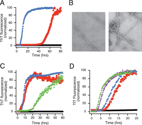
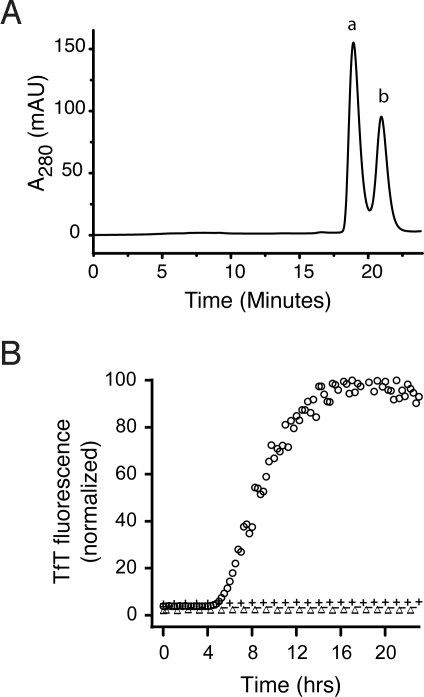
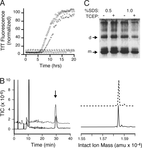
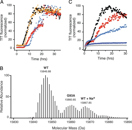
Familial amyotrophic lateral sclerosis (fALS) caused by mutations in copper-zinc superoxide dismutase (SOD1) is characterized by the presence of SOD1-rich inclusions in spinal cords. Similar inclusions observed in fALS transgenic mice have a fibrillar appearance suggestive of amyloid structure. Metal-free apo-SOD1 is a relatively stable protein and has been shown to form amyloid fibers in vitro only when it has been subjected to severely destabilizing conditions, such as low pH or reduction of its disulfide bonds. Here, by contrast, we show that a small amount of disulfide-reduced apo-SOD1 can rapidly initiate fibrillation of this exceptionally stable and highly structured protein under mild, physiologically accessible conditions, thus providing an unusual demonstration of a specific, physiologically relevant form of a protein acting as an initiating agent for the fibrillation of another form of the same protein. We also show that, once initiated, elongation can proceed via recruitment of either apo- or partially metallated disulfide-intact SOD1 and that the presence of copper, but not zinc, ions inhibits fibrillation. Our findings provide a rare glimpse into the specific changes in a protein that can lead to nucleation and into the ability of amyloid nuclei to recruit diverse forms of the same protein into fibrils.


Conflict of interest statement

The authors declare no conflict of interest.

Figures

Fig. 1.
Fig. 1.
Fibrillation of SOD1 can occur in mild conditions. Apo- SOD1 (50 μM) in 10 mM potassium phosphate buffer, pH 7.4 with additions as indicated was incubated with constant agitation at 37 °C in 96-well plates. TfT fluorescence was monitored as an indicator of fibril formation. (A) Fibrillation of SOD1 is stimulated by 1 M Gdm·HCl (red) or 50 mM DTT (blue). No addition is indicated by black circles. (B) Electron micrographs of SOD1 fibrils generated in the presence of 1 M Gdm·HCl (Left) and 5 mM DTT (Right). (Scale: 1 cm = 200 nm). (C) Multiple reducing agents catalyze fibril formation. Apo-SOD1 alone (black circles), with 10 mM DTT (red squares), 10 mM TCEP (blue triangles), or 10 mM glutathione (green triangles). (D) DTT concentration dependence of fibril formation for apo-SOD1. DTT concentrations are: 50 μM (black circles), 250 μM (red squares), 1 mM (blue triangles), 5 mM (purple inverted triangles), and 25 mM (green diamonds).
Fig. 2.
Fig. 2.
Disulfide-reduced SOD1 with free thiol groups initiates fibrillation in the absence of reducing agent. (A) Separation of SOD1SH-SH (peak b) and SOD1S-S (peak a) generated during the lag phase of a fibrillation reaction in 5 mM DTT performed in the manual format by SEC. (B) These peaks were used as isolated or treated with NEM to block free thiols and added to fibrillation reactions without reducing agent. Apo-SOD1 (47.5 μM) in phosphate buffer was incubated with 2.5 μM SOD1 in the following forms: SOD12SH (peak b), ○; SOD1S-S (peak 2), △; SOD1–4NEM (peak b, NEM treated), −; SOD1S-S-2NEM (peak a, NEM treated), +.
Fig. 3.
Fig. 3.
Both Cys-57 and Cys-146, but not Cys-6 or Cys-111, are necessary to initiate amyloid formation. (A) 2.5 μM SOD12SH (○), AS2SH (+), SOD1–4NEM (△), AS-2NEM (−), C57S (◇), and C146S (□) were added to 47.5 μM apo-SOD1 in the absence of reducing agents. (B) The amyloid nuclei elongate via noncovalent interactions to form mature fibrils. SOD1 fibrils grown in the presence of 5 mM DTT were denatured in 6.5 M Gdm·HCl with or without 20 mM TCEP and analyzed by HPLC-MS. (Left) Total ion count of Gdm·HCl-denatured (solid line) or Gdm·HCl-and-TCEP-denatured (dashed line) SOD1 fibrils. (Right) Reconstructed mass spectrum of peak at 30 min for LCMS of Gdm·HCl denaturation (solid line) and Gdm·HCl-TCEP denaturation (dashed line) reactions, showing monomeric SOD at 15,845 Da. (C) PAGE of amyloid fibrils grown in 5 mM DTT and denatured in SDS with or without 20 mM TCEP show similar amounts of monomers, dimers, and higher oligomers, suggesting fibril elongation does not involve intermolecular disulfide cross-linking.
Fig. 4.
Fig. 4.
Disulfide-reduced mutants can initiate fibrillation of SOD1, while copper, but not zinc, can inhibit fibril elongation. (A) Small amounts of disulfide-reduced fALS-SOD1 mutants can initiate fibril formation of apo-SOD1; 2.5 μM SOD12SH (red squares), G93A2SH (orange circles), G37R2SH (aqua triangles), and D101N2SH (black triangles) were incubated with 47.5 μM apo-SOD1 in the absence of reducing agents. Fibril formation was confirmed in all cases by electron microscopy (data not shown). (B) High-resolution mass spectrometry in an LTQ-FTMS instrument shows that WT SOD1 fibrils grown by the addition of G93A2SH and denatured in the presence of 6.5 M Gdm·HCl and 20 mM TCEP contain both WT SOD1 and G93A. Shown is the isotopically-resolved reconstructed intact-ion mass spectrum from the z = 10 peak. (C) Cu and Zn binding to apo-SOD1 can attenuate and, in some cases, abolish fibril formation initiated by SOD12SH; 2.5 μM SOD12SH was added to 47.5 μM apo-SOD1 (red squares), E2,EZn-SOD1 (black circles), as-isolated SOD1 (light blue triangles), Cu2E2-SOD1 (dark blue triangles), and Cu2Zn2-SOD1 (gray diamonds). As-isolated SOD1 refers to SOD1 purified from yeast. It contains 0.6 eq. copper and 2.5 eq. zinc ions per dimer.

Comment in

  • ALS precursor finally shaken into fibrils.
    Rousseau F, Schymkowitz J, Oliveberg M. Rousseau F, et al. Proc Natl Acad Sci U S A. 2008 Dec 2;105(48):18649-50. doi: 10.1073/pnas.0810568106. Epub 2008 Nov 24. Proc Natl Acad Sci U S A. 2008. PMID: 19033195 Free PMC article. No abstract available.

Similar articles

Cited by

References

    1. Valentine JS, Doucette PA, Potter SZ. Copper-zinc superoxide dismutase and amyotrophic lateral sclerosis. Annu Rev Biochem. 2005;74:563–593. - PubMed
    1. Tiwari A, Hayward LJ. Familial amyotrophic lateral sclerosis mutants of copper/zinc superoxide dismutase are susceptible to disulfide reduction. J Biol Chem. 2003;278:5984–5992. - PubMed
    1. Bruijn LI, Miller TM, Cleveland DW. Unraveling the mechanisms involved in motor neuron degeneration in ALS. Annu Rev Neurosci. 2004;27:723–749. - PubMed
    1. Kato S, et al. Pathological characterization of astrocytic hyaline inclusions in familial amyotrophic lateral sclerosis. Am J Pathol. 1997;151:611–620. - PMC - PubMed
    1. Shibata N, et al. Cu/Zn superoxide dismutase-like immunoreactivity in Lewy body-like inclusions of sporadic amyotrophic lateral sclerosis. Neurosci Lett. 1994;179:149–152. - PubMed

Publication types

LinkOut - more resources