Review
. 2008 Dec;138(12):2510-4.
doi: 10.3945/jn.108.096016.

Docosahexaenoic acid and the aging brain

Affiliations
Review

Docosahexaenoic acid and the aging brain

Walter J Lukiw et al. J Nutr. 2008 Dec.

Abstract

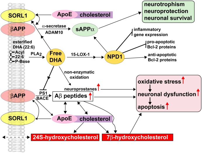
The dietary essential PUFA docosahexaenoic acid [DHA; 22:6(n-3)] is a critical contributor to cell structure and function in the nervous system, and deficits in DHA abundance are associated with cognitive decline during aging and in neurodegenerative disease. Recent studies underscore the importance of DHA-derived neuroprotectin D1 (NPD1) in the homeostatic regulation of brain cell survival and repair involving neurotrophic, antiapoptotic and antiinflammatory signaling. Emerging evidence suggests that NPD1 synthesis is activated by growth factors and neurotrophins. Evolving research indicates that NPD1 has important determinant and regulatory interactions with the molecular-genetic mechanisms affecting beta-amyloid precursor protein (betaAPP) and amyloid beta (Abeta) peptide neurobiology. Deficits in DHA or its peroxidation appear to contribute to inflammatory signaling, apoptosis, and neuronal dysfunction in Alzheimer disease (AD), a common and progressive age-related neurological disorder unique to structures and processes of the human brain. This article briefly reviews our current understanding of the interactions of DHA and NPD1 on betaAPP processing and Abeta peptide signaling and how this contributes to oxidative and pathogenic processes characteristic of aging and AD pathology.


Figures

FIGURE 1
FIGURE 1
DHA, NPD1, and βAPP-derived Aβ peptide signaling circuits in homeostatic aging and in AD. DHA and NPD1 act as PLA2- and 15-LOX-mediated neuroprotectants in the βAPP-sAPPα-Aβ peptide signaling pathway. Free DHA is liberated from membrane-bound stores via the action of a highly regulated membrane-associated PLA2 that may be subsequently converted into a potent neurotrophic NPD1 through an enzyme-mediated lipoxygenation via 15-LOX-1 or 15-LOX-like activities. NPD1 has been shown to convey multiple neuroprotective effects including induction of antiapoptotic Bcl-2 proteins, inhibition in the expression of proapoptotic Bcl-2 proteins, and suppression of inflammatory gene expression. Various ROS are more abundant in AD than in control brain, suggesting a possible role for oxidation-related decrease in protein function in processes such as depletion of the cellular redox balance, loss of specific protein function, interference with the cell cycle, and abnormal clearance of proteins and neurodegeneration leading ultimately to neuronal death. Nonenzymatic oxidation of free DHA results in the formation of neuroprostanes, a class of peroxidized lipids that further support oxidative stress, neuronal dysfunction, and apoptosis. Nonenzymatic reactions may be quenched by specific antioxidants and free radical scavengers, indicating that the redox state of brain cells has bearing on neurotrophic or oxidative-neurotoxic pathways for DHA. Enriched within neuronal plasma and endoplasmic membranes, the integral βAPP gives rise to sAPPα via an α-secretase/ADAM (a disintegrin and metalloprotease) 10-mediated pathway that is nonamyloidogenic and neurotrophic and whose synthesis is supported by free DHA and NPD1 (upper pathways). The βAPP membrane-integral sorting receptor sortilin-1 (SORL1), when proximal to βAPP, has direct effects on βAPP trafficking, and decreased abundance of SORL1, or βAPP-SORL1 dissociation, is coupled to activation of the amyloidogenic pathway from βAPP and the increased generation and secretion of Aβ peptides (lower pathways) (–49). SORL-1 further interacts with the type E apolipoprotein (ApoE), a major biolipid and cholesterol transporter in the brain, and the interaction of βAPP and ApoE within cholesterol-enriched lipid raft membrane domains, especially in the absence of SORL-1, gives rise to an increased generation of Aβ peptides via stimulation of β-amyloid cleavage enzyme (BACE) and presenilin 1 (PS1). The tandem actions of BACE and PS1 are sometimes referred to as the β-γ-secretase signaling pathway, an integral component of the amyloid cascade hypothesis, and known to contribute to Aβ peptide accumulation, neuropathology, and neurodegeneration. Aβ peptides bind directly to ApoE and cholesterol, and both Aβ peptides and βAPP oxidize ApoE-cholesterol to form the proapoptotic neurotoxic oxysterol 7β-hydroxycholesterol (7β-HC) or 24S hydroxycholesterol (24S-HC) via the action of CYP46A1 (–50). 24S-HC, highly enriched in the human CNS, is membrane permeable and is associated with amyloidogenesis and AD pathology (49). The actions of DHA or NPD1 on CYP46A1 and oxidation of cholesterol to 7β-HC or 24S-HC are not well understood. Current and emerging pharmaceutical strategies aim at the modulation of secretase activities through the actions of SALA to favor the more neurotrophic βAPP-cleavage signaling pathways (upper pathways) over the neurotoxic, amyloidogenic BACE-PS1 β-γ-secretase pathways (lower pathways; 50). The therapeutic use of statins, 3-hydroxy-3-methylglutaryl-coenzyme A reductase inhibitors, that lower serum cholesterol has also been shown to reduce Aβ peptide abundance in in vitro models of AD using human brain cell primary cultures and in some clinical trials, and large phase III studies are currently in progress (–50). The interactions of DHA and NPD1 with SALA drugs or statins are not well understood; however, early clinical trials using DHA and antioxidants together as enhancers of cognition in aged patients showed synergistic beneficial effects (23,49,50).

References

    1. Marszalek JR, Lodish HF. Docosahexaenoic acid, fatty acid-interacting proteins, and neuronal function. Annu Rev Cell Dev Biol. 2005;21:633–57. - PubMed
    1. Rapoport SI, Rao JS, Igarashi M. Brain metabolism of nutritionally essential polyunsaturated fatty acids depends on both the diet and the liver. Prostaglandins Leukot Essent Fatty Acids. 2007;77:251–61. - PMC - PubMed
    1. Innis SM. Dietary (n-3) fatty acids and brain development. J Nutr. 2007;137:855–9. - PubMed
    1. Spector AA. Plasma free fatty acid and lipoproteins as sources of polyunsaturated fatty acid for the brain. J Mol Neurosci. 2001;16:159–65. - PubMed
    1. Scott BL, Bazan NG. Membrane docosahexaenoate is supplied to the developing brain and retina by the liver. Proc Natl Acad Sci USA. 1989;86:2903–7. - PMC - PubMed

Publication types