. 2009 Feb;29(2):202-8.
doi: 10.1161/ATVBAHA.108.174391. Epub 2008 Nov 20.

GIT1 mediates VEGF-induced podosome formation in endothelial cells: critical role for PLCgamma

Affiliations

GIT1 mediates VEGF-induced podosome formation in endothelial cells: critical role for PLCgamma

Jing Wang et al. Arterioscler Thromb Vasc Biol. 2009 Feb.

Abstract

Objective: We and others showed that tyrosine kinase receptors (TKRs) such as the epidermal growth factor receptor stimulate G protein-coupled receptor (GPCR) kinase-interacting protein 1 (GIT1) phosphorylation via c-Src, which is required for phospholipase C-gamma (PLCgamma) activation, indicating that GIT1 participates in TKR signaling. VEGF is the most important TKR in endothelial cells (ECs); essential for cell survival, migration, and angiogenesis. Podosomes, actin-rich structures, were found to contribute to EC migration, tissue invasion, and matrix remodeling, suggesting a role for podosomes in angiogenesis. Because GIT1 is a substrate of c-Src, and podosome formation is c-Src dependent, we hypothesized that GIT1 plays an important role in VEGF-induced EC podosome formation and cell migration.

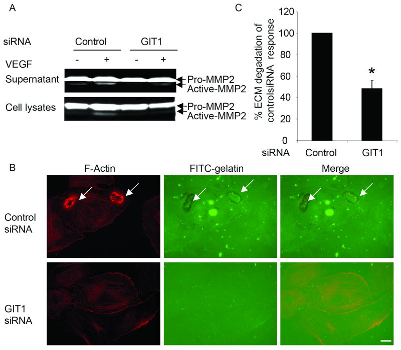
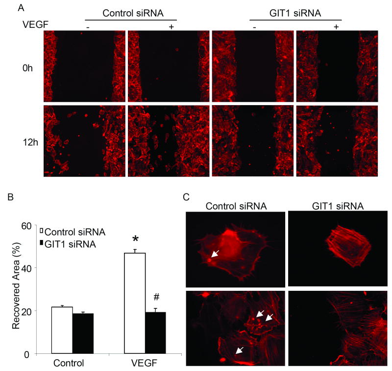
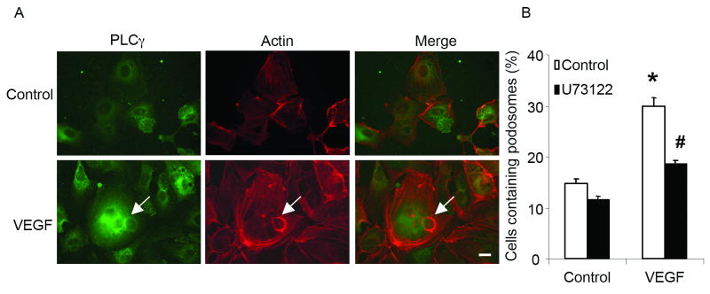
Methods and results: Exposure of ECs to VEGF for 30 minutes stimulated GIT1 colocalization with podosomes. Depletion of GIT1 by siRNA significantly decreased VEGF-induced podosome formation. A key role for PLCgamma was suggested by several experiments. Double staining PLCgamma and actin showed colocalization of PLCgamma with podosomes. Podosome formation was dramatically reduced by PLCgamma inhibitor U73122, Src inhibitor PP2, or expression of dominant negative small GTPases. Therefore, VEGF-induced EC podosome formation is dependent on Src, GIT1, PLCgamma, and small GTPases. In addition, matrix metalloprotease 2 (MMP2) and MT-MMP1 were detected at sites of VEGF-induced podosomes. Depletion of GIT1 by siRNA also significantly inhibited VEGF-induced MMP2 activation and extracellular matrix (ECM) degradation. Therefore, GIT1 mediates VEGF-induced matrix metalloproteinase (MMP) activation and ECM degradation by regulating podosome formation. Finally, depletion of GIT1 by siRNA significantly decreased VEGF-induced cell migration.

Conclusions: These data indicate that GIT1 is an essential mediator for VEGF-induced EC podosome formation and cell migration via PLCgamma.


Figures

Figure 1
Figure 1. GIT1 mediates VEGF-induced podosomes
(A) EC were transfected with 100 nM control siRNA or GIT1 siRNA using Lipofectamine 2000 for 48 hours, and cell lysates were immunoblotted with GIT1 antibody. Actin blot shows equal loading. (B) HUVEC were transfected with 100 nM siRNA as in (A), then treated with VEGF for varying times. Podosomes were identified as large phalloidin positive rings. Percentage of podosome positive cells (%) was determined. (*, P<0.05 vs control siRNA at time 0 min; #, P<0.05 vs control siRNA at 15 and 30 min) (C-K) HUVEC were seeded in 2% gelatin coated dishes. After cells were treated with 50 ng/ml VEGF for 30 min, cells were stained for GIT1 (Invitrogen, green) and F-actin (Sigma, red). Arrow shows podosomes. Bar represents 10 um.
Figure 2
Figure 2. PLCγ mediates VEGF-induced podosome formation
(A) Co-localization of PLCγ with podosomes. HUVEC were treated with 50 ng/ml VEGF for 30 min. Then cells were stained with PLCγ antibody (BD, green) and F-actin (Sigma, red). Arrow shows podosomes. Bar represents 10 um. (B) VEGF-induced podosome formation is dependent on PLCγ. HUVEC were pretreated with 5 μM U73122 for 30 min, then stimulated with 50 ng/ml VEGF. Percentage of podosome positive cells (%) was determined. (*, P<0.05 vs no VEGF treatment control; #, P<0.05 vs no U73312 treatment control).
Figure 3
Figure 3. VEGF-induced podosomes are Src and small GTPases dependent
(A) HUVEC were pretreated with 4-amino-5-(4-chlorophenyl)-7-(t-butyl)pyrazolo(3,4-d) pyrimidine (PP2) for 30 minutes, then stimulated with 50 ng/ml VEGF. Percentage of podosome positive cells (%) was determined. (*, P<0.05 vs no VEGF treatment control; #, P<0.05 vs no PP2 treatment control) (B) HUVEC were infected with Ad.DN small GTPases (Rho, RhoA, Cdc42) before treatment with VEGF, then cells were stained for podosomes. Percentage of podosome positive cells (%) was determined. (*, P<0.05 vs LacZ no VEGF treatment control; #, P<0.05 vs LacZ VEGF treatment control) (C) At the same time, cell lysates were used for western blotting to confirm the expression of adenovirus. Actin blots show equal loading.
Figure 4
Figure 4. Co-localization of MMP2 and MT1-MMP with podosomes
After treatment with 50 ng/ml VEGF for 30 minutes, cells were fixed and stained with MMP-2(A-F) or MT1-MMP (G-L) antibody (green), and TRITC-phalloidin (red). Arrows show podosomes. Bar represents 10 μm.
Figure 5
Figure 5. Role of GIT1 in MMP activity and ECM degradation
(A) HUVEC were transfected with 100 nM control siRNA or GIT1 siRNA, followed by treatment with 50ng/ml VEGF. Both concentrated supernatants and cell lysates were were analysed by 1% gelatin zymography. The active form of MMP-2 (lower molecular weight) indicates cleavage of pro-MMP2. (B) HUVEC were transfected with 100 nM control siRNA or GIT1 siRNA for 48 hours. Then, cells were seeded on FITC-gelatin-coated coverslips followed by treatment with 50 ng/ml VEGF. Cells were fixed and stained for F-Actin. Arrows show areas of degraded gelatin that overlap with podosomes. (C) Quantitative data were presented as percentages of control siRNA response obtained in the presence of VEGF. (*, P<0.05 vs control siRNA response)
Figure 6
Figure 6. GIT1 mediates VEGF-induced endothelial cell migration by affecting podosome formation
(A) After HUVEC were transfected with 100 nM control siRNA or GIT1 siRNA, scratch wound injury was performed with or without VEGF. (B) Migration rate was analyzed with NIH-Image software (*, p < 0.05 versus control siRNA at time 0hr, #, p< 0.05 versus control siRNA at time 12 hour). (C) Cells were fixed and stained with TRITC-phalloidin. Arrows show podosomes. Bar represents 10 μm.

References

    1. Neufeld G, Cohen T, Gengrinovitch S, Poltorak Z. Vascular endothelial growth factor (VEGF) and its receptors. Faseb J. 1999;13:9–22. - PubMed
    1. Werdich XQ, Penn JS. Src, Fyn and Yes play differential roles in VEGF-mediated endothelial cell events. Angiogenesis. 2005;8:315–326. - PubMed
    1. Abu-Ghazaleh R, Kabir J, Jia H, Lobo M, Zachary I. Src mediates stimulation by vascular endothelial growth factor of the phosphorylation of focal adhesion kinase at tyrosine 861, and migration and anti-apoptosis in endothelial cells. Biochem J. 2001;360:255–264. - PMC - PubMed
    1. Takahashi T, Yamaguchi S, Chida K, Shibuya M. A single autophosphorylation site on KDR/Flk-1 is essential for VEGF-A- dependent activation of PLC-gamma and DNA synthesis in vascular endothelial cells. Embo J. 2001;20:2768–2778. - PMC - PubMed
    1. Meyer RD, Latz C, Rahimi N. Recruitment and activation of phospholipase Cgamma1 by vascular endothelial growth factor receptor-2 are required for tubulogenesis and differentiation of endothelial cells. J Biol Chem. 2003;278:16347–16355. - PMC - PubMed

Publication types

MeSH terms