. 2009 Feb 5;113(6):1332-9.
doi: 10.1182/blood-2008-07-167148. Epub 2008 Nov 20.

Lactadherin and clearance of platelet-derived microvesicles

Affiliations

Lactadherin and clearance of platelet-derived microvesicles

Swapan K Dasgupta et al. Blood. .

Abstract

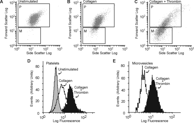
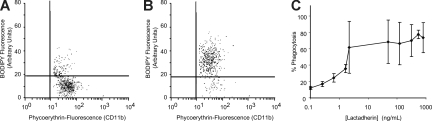
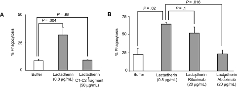
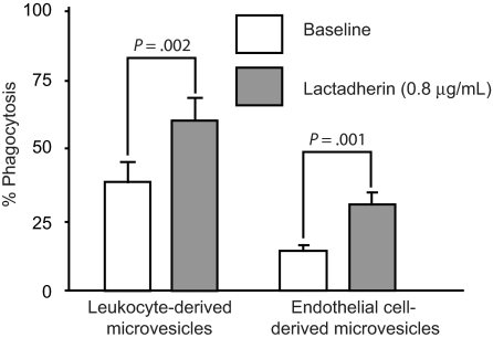
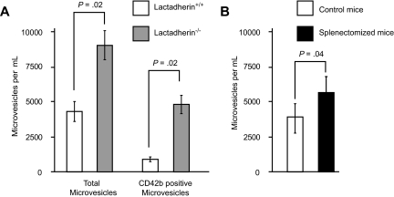
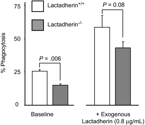
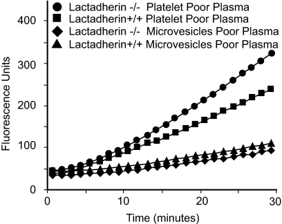
The transbilayer movement of phosphatidylserine from the inner to the outer leaflet of the membrane bilayer during platelet activation is associated with the release of procoagulant phosphatidylserine-rich small membrane vesicles called platelet-derived microvesicles. We tested the effect of lactadherin, which promotes the phagocytosis of phosphatidylserine-expressing lymphocytes and red blood cells, in the clearance of platelet microvesicles. Platelet-derived microvesicles were labeled with BODIPY-maleimide and incubated with THP-1-derived macrophages. The extent of phagocytosis was quantified by flow cytometry. Lactadherin promoted phagocytosis in a concentration-dependent manner with a half-maximal effect at approximately 5 ng/mL. Lactadherin-deficient mice had increased number of platelet-derived microvesicles in their plasma compared with their wild-type littermates (950 +/- 165 vs 4760 +/- 650; P = .02) and generated 2-fold more thrombin. In addition, splenic macrophages from lactadherin-deficient mice showed decreased capacity to phagocytose platelet-derived microvesicles. In an in vivo model of light/dye-induced endothelial injury/thrombosis in the cremasteric venules, lactadherin-deficient mice had significantly shorter time for occlusion compared with their wild-type littermate controls (5.93 +/- 0.43 minutes vs 9.80 +/- 1.14 minutes;P = .01). These studies show that lactadherin mediates the clearance of phosphatidylserine-expressing platelet-derived microvesicles from the circulation and that a defective clearance can induce a hypercoagulable state.


Figures

Figure 1
Figure 1
Lactadherin binding to platelets and platelet-derived microvesicles. Washed human platelets were treated with (A) buffer only, (B) collagen (50 μg/mL), or (C) a combination of thrombin (0.1 U/mL) and collagen (50 μg/mL), and FITC-lactadherin (5 μg/mL) and a PE-labeled anti-CD42b (2.5 μg/mL) were added. The generation of microvesicles was analyzed by flow cytometry as described before. To resolve the platelets and platelet-derived microparticles from background scatter, only CD42b+ events were analyzed for forward and side scattering. The gates for microvesicles (gate M) and intact platelets (gate P) were set with the use of isolated microvesicles and unstimulated platelets, respectively. Platelets and microvesicles were analyzed separately for the expression of phosphatidylserine by 5 μg/mL FITC-lactadherin (D,E).
Figure 2
Figure 2
Lactadherin is present in circulating platelet-derived microvesicles in normal human plasma. (A) Microvesicles, isolated from normal human plasma by centrifugation, were incubated with PE-labeled anti-CD42b (2.5 μg/mL) and FITC-antilactadherin antibody L688 (5 μg/mL) or an FITC-labeled irrelevant control antibody. The CD42b-expressing particles were gated and analyzed for FITC fluorescence. (B) Immunoblot of microvesicle-associated lactadherin. Protein A + G agarose beads (100 μL) were incubated overnight at 4°C with 100 μg rabbit polyclonal anti-IIb/IIIa antibody or an irrelevant rabbit control antibody. The beads were washed and incubated with 5 mL platelet-poor plasma and washed. Bound proteins were eluted in 50 μL 1% SDS, subjected to SDS-PAGE, transferred to PVDF membrane, and probed with the monoclonal antibody to lactadherin L688 and developed by a peroxidase-labeled goat anti–mouse antibody (1/2000 dilution) and chloronaphthol (0.3 mg/mL) and H2O2 (0.03%). (Lane 1) Anti-IIb/IIIa anti-body and (lane 2) irrelevant control antibody. (Lane 1) Immunoprecipitation with anti-IIb/IIIa and (lane 2) irrelevant control antibody.
Figure 3
Figure 3
Phagocytosis of platelet-derived microvesicles by macrophages. BODIPY-maleimide–labeled human platelet-derived microvesicles (80 μg) were incubated with THP-1–derived macrophages (106 cells/well) for 30 minutes. The nonadherent and the surface-bound microvesicles were detached with trypsin-EDTA solution. The macrophages were washed and analyzed by flow cytometry. The macrophages were identified by PE-labeled CD11b and gated. Phagocytosis was quantified by measuring the percentage of BODIPY (green) fluorescence-positive macrophages. (A) Phagocytosis in the absence of lactadherin. (B) Phagocytosis in the presence of lactadherin (0.8 μg/mL). (C) Lactadherin concentration-dependent phagocytosis of microvesicles. Each point represents the mean and SD of 3 or more separate experiments.
Figure 4
Figure 4
Inhibition of lactadherin-dependent phagocytosis of platelet-derived microvesicles. (A) THP-1–derived macrophages were incubated with BODIPY-labeled human platelet-derived microvesicles and lactadherin in the presence of C1C2 fragment (A) or abciximab (B). The extent of phagocytosis was measured as described in Figure 3. The results are the means and SDs of triplicate measurements.
Figure 5
Figure 5
Effect of lactadherin on the phagocytosis of leukocytes and endothelial cell–derived microvesicles. BODIPY-maleimide–labeled microvesicles from peripheral blood leukocytes or human umbilical vein endothelial cells were incubated with THP-1–derived macrophages (106 cells/well) for 30 minutes with or without lactadherin (0.8 μg/mL), and the extent of phagocytosis was quantified by flow cytometry by measuring the percentage of green fluorescence (BODIPY)–positive macrophages as in Figure 3.
Figure 6
Figure 6
Circulating microvesicles in mouse blood. (A) Plasma was collected from lactadherin-deficient mice and their littermate controls (N = 3 for each group). The microvesicles were quantified by flow cytometry, based on the light scatter and surface expression of CD42b (n = 3). (B) Wild-type mice were subjected to splenectomy, and the circulating microvesicles were measured 4 hours later. The results are the means and SDs (N = 3).
Figure 7
Figure 7
Phagocytosis of platelet-derived microvesicles by splenic macrophages in mice. Splenic macrophages were isolated from lactadherin-deficient mice and their wild-type littermate controls and incubated with BODIPY-labeled mouse platelet-derived microvesicles in the absence or presence of exogenous lactadherin. Phagocytosis of microvesicles was quantified as described before. The results are the means and SDs of triplicate measurements.
Figure 8
Figure 8
Thrombin generation in the plasma of lactadherin-deficient mice and control littermates. The reaction mixture consisted of plasma (80 μL) and the trigger solution (3 ng purified Russell viper venom in 40 μL), fluorogenic thrombin substrate Z-Gly-Gly-Arg-AMC (2.5 mM), and CaCl2 (10 mM). The generation of thrombin was measured as a function of fluorescence in a fluorimeter over a period of 30 minutes. Thrombin was then calculated using an α2-macroglobulin thrombin complex standard as described by Hemker et al. Representative data are presented. Blood samples from 5 mice from each group (lactadherin−/− and littermate controls [lactadherin+/+]) were pooled for each experiment.
Figure 9
Figure 9
Thrombus formation in lactadherin-deficient mice. Endothelial injury was induced by light/dye in vivo in the cremasteric venules. Thrombus onset and flow cessation were monitored by intravital microscopy in lactadherin-deficient mice (formula image
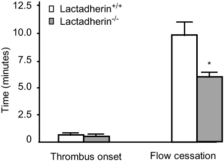
) and the littermate controls (□). *P = .01; n = 9.

References

    1. Zwaal RF, Comfurius P, Bevers EM. Surface exposure of phosphatidylserine in pathological cells. Cell Mol Life Sci. 2005;62:971–988. - PMC - PubMed
    1. Bevers EM, Comfurius P, Zwaal RF. Regulatory mechanisms in maintenance and modulation of transmembrane lipid asymmetry: pathophysiological implications. Lupus. 1996;5:480–487. - PubMed
    1. Zwaal RF, Schroit AJ. Pathophysiologic implications of membrane phospholipid asymmetry in blood cells. Blood. 1997;89:1121–1132. - PubMed
    1. Solum NO. Procoagulant expression in platelets and defects leading to clinical disorders. Arterioscler Thromb Vasc Biol. 1999;19:2841–2846. - PubMed
    1. Comfurius P, Senden JM, Tilly RH, Schroit AJ, Bevers EM, Zwaal RF. Loss of membrane phospholipid asymmetry in platelets and red cells may be associated with calcium-induced shedding of plasma membrane and inhibition of aminophospholipid translocase. Biochim Biophys Acta. 1990;1026:153–160. - PubMed

Publication types

MeSH terms