. 2008 Nov;4(11):e1000217.
doi: 10.1371/journal.ppat.1000217. Epub 2008 Nov 21.

the hyphal-associated adhesin and invasin Als3 of Candida albicans mediates iron acquisition from host ferritin

Affiliations

the hyphal-associated adhesin and invasin Als3 of Candida albicans mediates iron acquisition from host ferritin

Ricardo S Almeida et al. PLoS Pathog. 2008 Nov.

Abstract

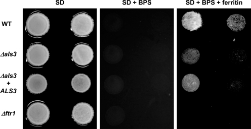
Iron sequestration by host iron-binding proteins is an important mechanism of resistance to microbial infections. Inside oral epithelial cells, iron is stored within ferritin, and is therefore not usually accessible to pathogenic microbes. We observed that the ferritin concentration within oral epithelial cells was directly related to their susceptibility to damage by the human pathogenic fungus, Candida albicans. Thus, we hypothesized that host ferritin is used as an iron source by this organism. We found that C. albicans was able to grow on agar at physiological pH with ferritin as the sole source of iron, while the baker's yeast Saccharomyces cerevisiae could not. A screen of C. albicans mutants lacking components of each of the three known iron acquisition systems revealed that only the reductive pathway is involved in iron utilization from ferritin by this fungus. Additionally, C. albicans hyphae, but not yeast cells, bound ferritin, and this binding was crucial for iron acquisition from ferritin. Transcriptional profiling of wild-type and hyphal-defective C. albicans strains suggested that the C. albicans invasin-like protein Als3 is required for ferritin binding. Hyphae of an Deltaals3 null mutant had a strongly reduced ability to bind ferritin and these mutant cells grew poorly on agar plates with ferritin as the sole source of iron. Heterologous expression of Als3, but not Als1 or Als5, two closely related members of the Als protein family, allowed S. cerevisiae to bind ferritin. Immunocytochemical localization of ferritin in epithelial cells infected with C. albicans showed ferritin surrounding invading hyphae of the wild-type, but not the Deltaals3 mutant strain. This mutant was also unable to damage epithelial cells in vitro. Therefore, C. albicans can exploit iron from ferritin via morphology dependent binding through Als3, suggesting that this single protein has multiple virulence attributes.


Conflict of interest statement

The authors have declared that no competing interests exist.

Figures

Figure 1
Figure 1. The ferritin content of host epithelial cells influences the cell damage by C. albicans.
The ferritin content was monitored using immunofluorescence (red, antibody staining for ferritin; blue, nuclei staining with DAPI). Following the treatments described in (A), (B) and (C), the monolayers were washed and incubated for 8 h in serum-free RPMI with 106 iron starved C. albicans (SC5314) cells. Cell damage was quantified by monitoring the release of epithelial LDH into the medium. (A) Monolayer incubated for 24 h in serum-free RPMI with 50 µM BPS. (B) Monolayer incubated 24 h in RPMI with 10% FBS (control). (C) Monolayer incubated for 24 h in RPMI with 10% FBS and 50 µM iron chloride. Bar indicates 10 µm. (D) Cell damage caused by C. albicans, calculated in relative cytotoxicity (%). Control, monolayers preincubated in normal cell culture medium (RPMI 1640 medium with 10% FBS); BPS, monolayers preincubated in serum-free RPMI with 50 µM BPS (iron chelator); Fe20, Fe30 and Fe50, monolayers preincubated in cell culture medium with 20, 30 and 50 µM ferric iron respectively. The experiment was performed twice in duplicates. *, significant difference compared to the control (p<0.05).
Figure 2
Figure 2. Invasion of ferritin depleted or enriched epithelial cells by C. albicans.
Approximately 105 iron starved C. albicans cells (SC5314) were co-incubated with ferritin depleted (BPS), ferritin enriched (Fe50) or non-treated (Control ) epithelial cells for 3 h. After fixation the samples were differentially stained and analyzed under the fluorescence microscope. The experiment was performed three times in duplicate. *, significant difference compared to non-treated epithelial cells (control) (p<0.001).
Figure 3
Figure 3. Usage of ferritin by C. albicans requires the reductive pathway and is mediated by acidification of the medium.
(A) SD agar plates were adjusted to pH 7.4 with 25 mM HEPES buffer and incubated for 3 days at 37°C under 5% CO2 (Ca, C. albicans SC5314) or 30°C without CO2 (Sc, S. cerevisiae ATCC9763). Iron indicates 50 µM iron sulphate. Ferritin indicates 20 µg/ml ferritin. Hemoglobin indicates 20 µg/ml hemoglobin. (B) C. albicans wild-type (SC5314) cells were spotted on YNB agar with the addition of either glucose (SD) or casamino acids as a carbon source and buffered with 25 or 200 mM HEPES. BPS (iron chelator) was used to remove free iron from the media. The growth of C. albicans strains and S. cerevisiae on agar with different iron sources was repeated at least 3 times.
Figure 4
Figure 4. Ferritin binding of C. albicans requires hyphal formation.
(A) C. albicans wild-type and mutant cells lacking key genes required for hyphal formation were incubated under hyphal-inducing conditions (RPMI 1640 medium, 37°C with 5% CO2) for 3 h. After 1 h in the presence of 100 µg/ml ferritin, cells were washed and ferritin was stained using immunofluorescence. Note that ferritin binding does not occur on the mother cell of hyphae. DIC, Differential Interference Contrast. Bar indicates 10 µm. (B) Quantification of C. albicans wild-type and mutant cells binding ferritin. For each strain the % ferritin binding cells is given for >100 randomly selected cells. The experiment was performed at least 3 times in duplicate. (C) Wild-type cells binding ferritin were analyzed under transmission electron microscopy. The black arrow points to the cell wall, the white arrow to ferritin molecules visualized by their electron density.
Figure 5
Figure 5. Transcription profiling identifies genes associated with ferritin binding.
To identify genes necessary for ferritin binding, C. albicans wild-type, Δhgc1 and Δras1 mutant cells were incubated under conditions which favored ferritin binding to wild-type and Δhgc1, but not Δras1 cells. RNA of each population of cells was isolated and used for microarray analysis. The micrographs show representative anti-ferritin labeled cells at the time point of RNA isolation. The Venn diagram indicates the number of genes up-regulated in wild-type and Δhgc1 and either unchanged or down-regulated in Δras1, as compared to a common control. Twenty two genes were up-regulated in wild-type and Δhgc1, but not Δras1 cells as expected for a ferritin receptor. Microarray experiments were performed in four biological replicates (two of them using dye swap). Note that the schematic presentation of the Venn diagram combines up-regulated (wild-type and Δhgc1) and unaltered or down-regulated (Δras1) genes to clarify the selection strategy.
Figure 6
Figure 6. Als3 is essential for ferritin binding.
(A) Mutants lacking either ALS3, HYR1 or ECE1–the three selected genes predicted to encode ferritin receptors–were tested for ferritin binding. Bar indicates 10 µm. (B) Ferritin binding was quantified by counting >100 randomly selected cells using fluorescence microscopy. *, significant difference compared to wild-type (p<0.0001). (C) Ferritin binding by mutants lacking key regulators of ALS3 expression (Δtec1 and Δbcr1). Bar indicates 10 µm.
Figure 7
Figure 7. Flow cytometric detection of ferritin binding.
C. albicans cells were incubated under hyphal-inducing conditions (RPMI, 37°C with 5% CO2) for 2 h. After 1 h in the presence of 100 µg/ml ferritin, cells were washed and ferritin was stained using indirect immunofluorescence and then analyzed using flow cytometry. (A) wild-type (CAF2-1); (B) Δals3; (C) Δals3+ALS3. Fluorescence data for 10,000 cells of each strain were collected. (D) Binding quantification. The data are expressed as a percentage of the results obtained with the wild-type strain (CAF2-1). The experiment was performed twice in duplicate. *, significant difference compared to wild-type (p<0.002).
Figure 8
Figure 8. Binding is necessary for iron acquisition from ferritin.
C. albicans wild-type (CAF2-1), Δals3 and Δftr1 were grown on media containing ferritin as the sole source of iron. SD agar was buffered using 100 mM HEPES (pH 7.4). BPS, iron chelator; ferritin, 2 µg/ml ferritin. Cells were spotted at two concentrations (left to right, 105 and 104 cells, respectively) for each strain. All plates were incubated for 3 days at 37°C under 5% CO2. The assay was performed three times.
Figure 9
Figure 9. Als3 is a ferritin receptor.
S. cerevisiae cells overexpressing ALS1, ALS3, ALS5 (driven by the ADH promoter) or carrying an empty plasmid (pADH) were incubated for 15 min in the presence of 25 µg/ml ferritin coupled to a fluorescent dye. Cells were washed to remove non-bound ferritin and analyzed with fluorescence microscopy in duplicate repeated three times. Bar indicates 10 µm.
Figure 10
Figure 10. C. albicans hyphae invading oral epithelial cells bind ferritin.
C. albicans wild-type (SC5314), Δals3 mutant and Δals3+ALS3 re-integrant cells were co-incubated with ferritin-enriched oral epithelial cells and differentially stained. (A), (E), (I) and (M); staining of extracellular (non-invaded) C. albicans with concanavalin A conjugated with fluorescein before cell permeabilization. (B), (F), (J) and (N); calcofluor white staining of whole C. albicans cells following epithelial cell permeabilization. (C), (G), (K) and (O); fluorescent dye (DY649) coupled antibody staining of ferritin. White arrows indicate hyphae surrounded by epithelial ferritin. (D), (H), (L) and (P); merged images. Bar in (P) indicates 10 µm.
Figure 11
Figure 11. Iron uptake from ferritin plays a role in oral epithelial cell damage.
C. albicans wild-type (CAF2-1), Δals3 mutant and Δftr1 mutant cells were co-incubated with oral epithelial cells. The monolayers were incubated for 8 h in serum-free RPMI 1640 with 106 C. albicans cells and cell damage was quantified by monitoring the release of epithelial LDH into the medium. The experiment was performed five times in triplicate. *, significant difference compared to the wild-type (p<0.0001).
Figure 12
Figure 12. Proposed model for iron utilization from ferritin by C. albicans.
Ferritin is a novel iron source used by C. albicans. In its hyphal form, C. albicans binds ferritin using Als3. Acidification of the surrounding environment mediates iron release from the ferritin shell and the released iron is then transported into the cell via the reductive pathway.

References

    1. Weinberg ED. Iron loading and disease surveillance. Emerg Infect Dis. 1999;5:346–352. - PMC - PubMed
    1. Andrews SC, Robinson AK, Rodriguez-Quinones F. Bacterial iron homeostasis. FEMS Microbiol Rev. 2003;27:215–237. - PubMed
    1. Howard DH. Iron gathering by zoopathogenic fungi. FEMS Immunol Med Microbiol. 2004;40:95–100. - PubMed
    1. Ekins A, Khan AG, Shouldice SR, Schryvers AB. Lactoferrin receptors in gram-negative bacteria: insights into the iron acquisition process. Biometals. 2004;17:235–243. - PubMed
    1. Gerlach GF, Anderson C, Potter AA, Klashinsky S, Willson PJ. Cloning and expression of a transferrin-binding protein from Actinobacillus pleuropneumoniae. Infect Immun. 1992;60:892–898. - PMC - PubMed

Publication types