. 2008 Nov;4(11):e1000211.
doi: 10.1371/journal.ppat.1000211. Epub 2008 Nov 21.

Interrelationship between dendritic cell trafficking and Francisella tularensis dissemination following airway infection

Affiliations

Interrelationship between dendritic cell trafficking and Francisella tularensis dissemination following airway infection

Erez Bar-Haim et al. PLoS Pathog. 2008 Nov.

Abstract

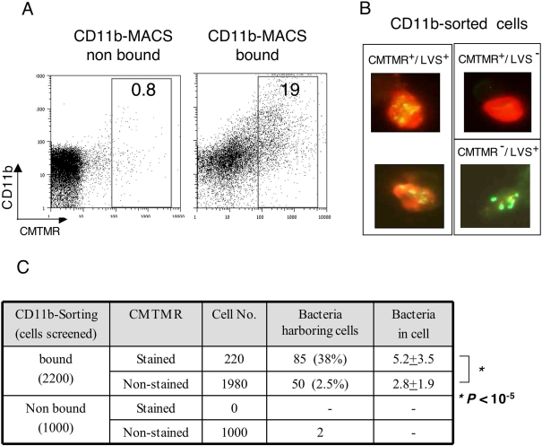
Francisella tularensis, the etiological agent of the inhalation tularemia, multiplies in a variety of cultured mammalian cells. Nevertheless, evidence for its in vivo intracellular residence is less conclusive. Dendritic cells (DC) that are adapted for engulfing bacteria and migration towards lymphatic organs could serve as potential targets for bacterial residence and trafficking. Here, we focus on the in vivo interactions of F. tularensis with DC following airway infection of mice. Lethal airway infection of mice with the live vaccine strain (LVS) results in trafficking of a CD11b(high)/CD11c(med)/autofluorescence(low) DC subset from the respiratory tract to the draining mediastinal lymph node (MdLN). Simultaneously, a rapid, massive bacterial colonization of the MdLN occurs, characterized by large bacterial foci formation. Analysis of bacteria in the MdLN revealed a major population of extracellular bacteria, which co-exists with a substantial fraction of intracellular bacteria. The intracellular bacteria are viable and reside in cells sorted for DC marker expression. Moreover, in vivo vital staining experiments indicate that most of these intracellular bacteria ( approximately 75%) reside in cells that have migrated from the airways to the MdLN after infection. The correlation between DC and bacteria accumulation in the MdLN was further demonstrated by manipulating DC migration to the MdLN through two independent pathways. Impairment of DC migration to the MdLN, either by a sphingosine-1-phosphate receptor agonist (FTY720) or by the D prostanoid receptor 1 agonist (BW245C), resulted in reduced bacterial colonization of MdLN. Moreover, BW245C treatment delayed the onset of morbidity and the time to death of the infected mice. Taken together, these results suggest that DC can serve as an inhabitation niche for F. tularensis in the early stages of infection, and that DC trafficking plays a role in pathogen dissemination. This underscores the therapeutic potential of DC migration impairing drugs in tularemia treatment.


Conflict of interest statement

The authors have declared that no competing interests exist.

Figures

Figure 1
Figure 1. Interaction of BMDC and LVS in vitro Alter.
(A) Propagation of LVS in J774A.1 cells and BMDC: Infection was performed as described in the Materials and Methods section, and gentamicin was added 1 hr post pulsing with bacteria. Cells were harvested 2 or 24 hrs post infection, washed, and lysed with DOC. Lysates were diluted and plated for CFU enumeration. Each bar represents the average CFUs in three infection wells. The entire experiment represents one of three repetitions. For visualization of cell-associated bacteria by fluorescence microscopy, infected J774A.1 or BMDC cells were stained with anti-LVS antibodies 24 hrs post LVS infection. (B) Expression of CCR7 by BMDC: Cells were analyzed for CCR7 expression 24 hours post infection with the live or formaldehyde-killed LVS. Non-pulsed cells and cells pulsed with 1 µg/ml E. coli LPS served as negative and positive controls, respectively. Gray lines denote isotype-matched immunoglobulin controls. Fractions of CCR7positive cells are indicated. (C) DC were pulsed as indicated above, and expression of co-stimulatory molecules was examined as described in Materials and Methods. Geomeans of fluorescence intensities are presented as relative values, using the values obtained for each marker by pulsing with 1 µg/ml E. coli LPS as 100. Results are presented by bar diagrams as the averages of three independent experiments. (D) Migration of BMDC: Cells were pulsed as indicated above, 24 hrs later cells were examined for migration in Transwell chambers towards CCL19 or control medium. Non-pulsed cells and cells pulsed with LPS served as controls. Results are presented as percent cells migrating from the upper compartments to the lower compartment. Each bar represents the average migration in 3 Transwells. The experiment was repeated 3 times. Data presented in this figure were generated by using BALB/c BMDC. Results were confirmed with DC derived from C57BL6 mice.
Figure 2
Figure 2. Spreading of LVS in intranasally infected mice.
Mice were infected intra-nasally with 105 CFU of LVS. (A) Bacteria were counted in BALF, lung tissue after lavage, MdLNs, spleens, and livers in animals sacrificed at the indicated time points after infection. Single cell suspensions derived from the various organs were diluted in PBS and plated for CFU counts. Each bar represents the average counts in 3 individual animals. Experiment was repeated 2 more times, resulting in similar observations. (B) Microscopy evaluation of MdLN colonization was performed on cryo-sections of MdLN, 72 hrs post infection with LVS. Sections were stained with rabbit anti-LVS hyperimmune serum and FITC-conjugated secondary antibody. One representative large infection focus is shown.
Figure 3
Figure 3. Definition of cell phenotypes in infected lungs and MdLNs.
Single cell suspensions of MdLN (A), washed lung (B), and BALF (C) were prepared from organs collected 48 hrs post infection with 105 CFU of LVS. Cells derived from non-infected animals served as controls. MdLN and BALF cell suspensions were stained for expression of CD11c and CD11b using antibodies conjugated to APC or PE, and analyzed by flow cytometry for surface display of markers as well as autofluorescence at FL1. Data are presented in the form of dot diagrams using parallel gating for identification of infection-triggered recruitments (numbers indicate percentage representations of the gated populations). Isotype-matched antibodies served as controls, and the same gates used in specific stain analysis were evaluated. In this case, gated population frequencies in infected organs were comparable to backgrounds (not shown). All analyses presented were conducted on tissues pooled from 3 individual animals. Three independent experiments exhibited similar cell profiles.
Figure 4
Figure 4. Cell recruitment to MdLN following airway infection with LVS.
MdLNs were isolated at various time points after intra-nasal infection of mice. Single cell suspensions were analyzed by flow cytometry as indicated on the legend to Figure 3. Number of AMΦ and RTDC at different time points post infection are presented as bar diagrams.
Figure 5
Figure 5. Identification of cells immigrating from airways to the MdLN by in vivo viable staining.
Airway cells of live mice were stained by instillation of CMTMR. Migration to lymph node was induced by intranasal infection with 107 CFU of LVS, using PBS instillation as control. Single cell suspensions were prepared from a pool of 8 lymph nodes collected 18 hrs post infection. Cells were analyzed by flow cytometry prior to (top panel, A and B) or after sorting by MACS with CD11b-microbeads (lower panel, C–F). Cells were stained with either APC-conjugated anti-CD11c (A,B,D,F) or APC conjugated anti-CD11b (C,E). Surface marker staining was plotted against CMTMR staining. Data are presented as dot diagrams. Gating for CMTMR staining and marker staining was based on diagrams of the appropriate non-stained controls (not shown). Numbers indicate cell percentage of the gated populations or of that defined by quadrants. Similar results were obtained in two other similar experiments.
Figure 6
Figure 6. Association of LVS with a specific cell population in infected lymph nodes.
Mice were infected intra-nasally with 105 CFU of LVS. Forty-eight hours later MdLNs were collected from 10 animals and single cell suspensions were prepared and examined by several assays. (A) Association between viable bacteria and MdLN cells was assayed by examining non-washed (total LN suspension) and washed cells for presence of viable LVS prior or post gentamicin treatment. Bacterial counts are presented as CFU per a single MdLN. (B) Association between LVS and RTDC cells was assayed by subjecting the washed cell fraction (∼108 cells) to sorting by magnetic microbeads coated with either anti-CD11b (B, top panel) or anti-CD11c (B, lower panel). Viable bacteria (no gentamicin treatment) were counted in the input cell suspension, in the bound cell fraction, as well as in cells that did not bind to the microbeads, and presented as CFU/106 cells. The amounts of cells in the input and in the bound fractions were ∼108/10 MdLNs and in the bound fractions only ∼106/10 MdLNs. CD11b and CD11c sorted cells were also examined by fluorescence microscopy for presence of intracellular bacteria. Typical cells carrying bacteria are presented in the insets to (B). (C) The cell/bacteria association in the CD11b-sorted fraction was characterized by determining bacterial count in non-treated cells, in cells treated with gentamicin (genta), in cells treated with gentamicin followed by washing and saponin lysis (genta+sap), as well as in cells where saponin treatment preceded gentamicin treatment (sap+genta). All bacterial counts were performed in triplicates, error bars represent variation of these counts. Data presented in this figure were collected from a single experiment. Two additional CD11b sorting experiments and one additional CD11c sorting experiment were conducted, exhibiting similar enrichment of intracellular bacteria.
Figure 7
Figure 7. Association of LVS with newly immigrating cells in infected lymph nodes.
Airway cells of live mice were stained by instillation of CMTMR. Migration to lymph node was induced by intranasal infection with 105 CFU of LVS. Single cell suspensions were prepared from pools of 8 lymph nodes, collected 48 hrs post infection. Cells were sorted by MACS with CD11b-microbeads. Bound and non bound cells were analyzed by flow cytometry (A) to evaluate the fraction of CMTMR+/CD11b+ cells. Sorted cells were examined by florescence microscopy for presence of the indicated cell phenotype (B). Large numbers of cells were screened to determine the proportion of the various cell populations (C). In addition, the average number of bacteria per cell in LVS-bearing CMTMR+ and CMTMR cells was determined (C, last column).
Figure 8
Figure 8. The effects of FTY720 on accumulation of RTDC and LVS in the MdLN.
Mice infected with 105 LVS were treated with FTY720 diluted in carrier solution, or with carrier solution alone, as described in Materials and Methods. To examine DC trafficking, MdLNs were collected 48 hrs post infection and analyzed for RTDC representation (CD11bhigh/autofluorescencelow) as described in the legend to Figure 3. An experiment was conducted on MdLN pools, each consisting of 6 organs. MdLNs were derived from infected mice treated with either carrier alone (A) or with FTY720 (B). Non-infected, FTY720-treated mice (C) served as controls. Bacterial colonization of the MdLN (D) was examined 48 hrs post infection in each one of the 6 individual FTY720- treated and 6 mock-treated mice, P<0.01. Three independent experiments, each conducted with groups of six mice, exhibited similar correlation between RTDC and LVS recruitment.
Figure 9
Figure 9. The effects of BW245C on the outcome of LVS infection.
Mice infected with 105 LVS were treated with BW245 diluted in carrier, or with carrier alone, as described in Materials and Methods. To examine DC trafficking (A), MdLNs were collected 48 hrs post infection and analyzed for RTDC representation (CD11bhigh/autofluorescencelow). Experiments were conducted on MdLN pools, each consisting of 6 organs, derived from infected or non-infected mice treated with either BW245C or carrier alone. Results are presented by bar diagrams as the averages of three independent experiments. * P<0.05. Bacterial colonization of the MdLN (B) was examined 48 hrs post infection in 13 individual BW245C-treated and 13 mock-treated mice. ** P<0.05. The effect of BW245 on mortality (C) was examined in groups of 10 mice comparing treated (open squares) and mock-treated mice (closed circles). The experiment presented represents one of three similar experiments. The effect of the drug on morbidity (D) was examined by monitoring weight loss for 5 consecutive days in groups of 10 mice each (8-week-old females). Groups consisted of non-infected BW245-treated mice (closed triangles), infected and treated mice (open squares), and infected mock-treated mice (closed circles). Data are presented as percentage of the weight determined on day zero and calculated separately for each individual mouse. *** P-values on days 2–5 range between <0.01 and <0.05.

References

    1. Steinman RM, Hemmi H. Dendritic cells: translating innate to adaptive immunity. Curr Top Microbiol Immunol. 2006;311:17–58. - PubMed
    1. Forster R, Schubel A, Breitfeld D, Kremmer E, Renner-Muller I, et al. CCR7 coordinates the primary immune response by establishing functional microenvironments in secondary lymphoid organs. Cell. 1999;99:23–33. - PubMed
    1. Hintzen G, Ohl L, del Rio ML, Rodriguez-Barbosa JI, Pabst O, et al. Induction of tolerance to innocuous inhaled antigen relies on a CCR7-dependent dendritic cell-mediated antigen transport to the bronchial lymph node. J Immunol. 2006;177:7346–7354. - PubMed
    1. Jakubzick C, Tacke F, Llodra J, van Rooijen N, Randolph GJ. Modulation of dendritic cell trafficking to and from the airways. J Immunol. 2006;176:3578–3584. - PubMed
    1. Hammad H, de Heer HJ, Soullie T, Hoogsteden HC, Trottein F, et al. Prostaglandin D2 inhibits airway dendritic cell migration and function in steady state conditions by selective activation of the D prostanoid receptor 1. J Immunol. 2003;171:3936–3940. - PubMed

Publication types