Review
. 2008 Dec;8(12):1999-2014.
doi: 10.1039/b811314a. Epub 2008 Oct 29.

Towards non- and minimally instrumented, microfluidics-based diagnostic devices

Affiliations
Review

Towards non- and minimally instrumented, microfluidics-based diagnostic devices

Bernhard Weigl et al. Lab Chip. 2008 Dec.

Abstract

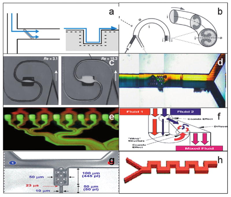
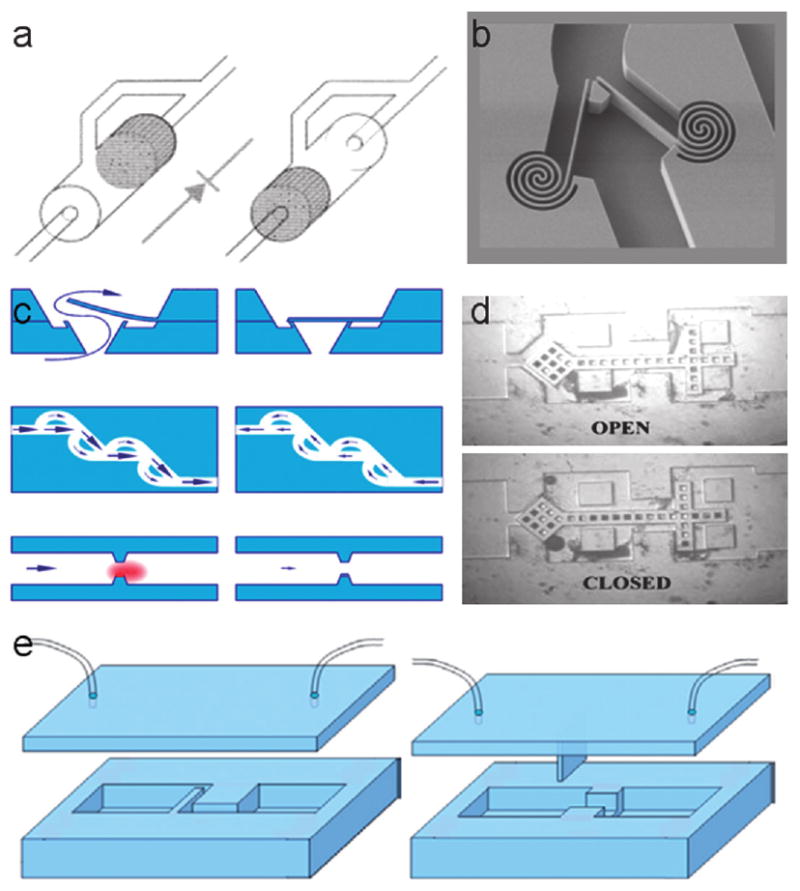
In many health care settings, it is uneconomical, impractical, or unaffordable to maintain and access a fully equipped diagnostics laboratory. Examples include home health care, developing-country health care, and emergency situations in which first responders are dealing with pandemics or biowarfare agent release. In those settings, fully disposable diagnostic devices that require no instrument support, reagent, or significant training are well suited. Although the only such technology to have found widespread adoption so far is the immunochromatographic rapid assay strip test, microfluidics holds promise to expand the range of assay technologies that can be performed in formats similar to that of a strip test. In this paper, we review progress toward development of disposable, low-cost, easy-to-use microfluidics-based diagnostics that require no instrument at all. We also present examples of microfluidic functional elements--including mixers, separators, and detectors--as well as complete microfluidic devices that function entirely without any moving parts and external power sources.


Figures

Fig. 1
Fig. 1
Schematic of immunochromatographic strip (ICS) test for gonorrhoea specifically developed for low-resource settings.
Fig. 2
Fig. 2
Example of a finger bellows that primes a wicking pad that then takes over the pumping action.
Fig. 3
Fig. 3
Springfusor, an example of a simple, portable, spring-loaded infusion pump.
Fig. 4
Fig. 4
Configuration of HydroLogic technology. A pressure signal received in the thrust inlet pressurizes the pressure capsule, which in turn causes the acidic solution to pressurize. The pressurized acidic solution ruptures the barrier to the alkaline cell. Microencapsulation of the alkaline controls the delay of a gas-generating reaction and its rate, providing means for controlling the subsequent events in the HydroLogic system.
Fig. 5
Fig. 5
Spiral polymerase chain reaction channel designed by PATH.
Fig. 6
Fig. 6
Front (a) and back (b) of exothermal circulation polymerase chain reaction card developed by Wheeler et al. The authors have designed a version of this device that would rely on exothermic heating materials instead of electric heaters, thus making the device non-instrumented.
Fig. 7
Fig. 7
Diagrams of various passive mixing methods: (a) transverse mixing wells, (b) spiral microchannels, (c) expansion vortex, (d) channel obstacles (e) lamination split and recombine, (f) Tesla split and recombine, (g) hybrid (obstacle nozzle), and (h) square wave mixer.
Fig. 8
Fig. 8
Examples of check valves include: (a) an on-chip check valve and diverter valve described by Hasselbrink, (b) a spring check valve design, (c) a type of PDMS “flap and block” described by Yang et al., (d) a free-floating check valve characterized by Deshmukh, and (e) a design for a flap check valve.
Fig. 9
Fig. 9
Schematic of a separation method relying upon microfluidic/micromagnetic forces to selectively remove living cells from flowing biological fluids without any wash steps.
Fig. 10
Fig. 10
A prototype device designed to collect 100 μL of blood from a fingerstick and transfer diluted blood to a microfluidic diagnostic disposable.
Fig. 11
Fig. 11
Easy-to-use, fast, and easy-to-interpret disposable device for ABO blood typing developed by Micronics, Inc. All fluids are moved and aliquoted through capillary force and manual on-card bellows pumping. Reagents and sample are mixed passively along laminar flow diffusion interfaces in microchannels. The result visible in the viewing window indicates blood type A, Rh positive.
Fig. 12
Fig. 12
Hydrostatically driven, integrated T-sensor design (a). A sample is put into the top left reservoir, a reagent (e.g., an indicator dye) is put into the top middle reservoir, and a reference solution with a known concentration of analyte is put into the right reservoir. The comparison of the intensity and position of the two diffusion interaction zones allows a semiquantitative analyte determination. The photograph in the center (b) shows such a T-sensor in operation, standing vertical, as it determines the pH of a buffer solution.
Fig. 13
Fig. 13
Hydrostatic-pressure-driven, integrated H-filter design. The figure shows an H-filter experiment in progress (illuminated with a UV lamp). A sample (e.g., blood) is put into the top left reservoir, and an acceptor reagent (e.g., water or saline) is put into the top right reservoir. Two parallel laminar streams will flow. Smaller components of the sample stream will diffuse into the acceptor stream. The two parallel flows are then split into two separate reservoirs (at the bottom of the drawing) that are connected only through a common vent. The left reservoir then contains the sample solution with a reduced concentration of the extracted component, and the right reservoir contains the acceptor reagent containing the extracted reagent. Both reservoirs can then be harvested from the cartridge for further use or be processed through further integrated microfluidic structures.
Fig. 14
Fig. 14
Prototype filter dial device for an integrated sample processing device with on-board reagents. Reverse transcription mix stored in two detachable vials: (1) POC, for emerging point-of-care NAT technologies and (2) CF, for central facility surveillance testing.
Fig. 15
Fig. 15
Diagram of how a PCM can be used to control temperature.
Fig. 16
Fig. 16
Thermal profile measured from a modified commercially available self-heating beverage product. Time is in minutes. Although the correct temperature range was not achieved for LAMP, the data demonstrates the capacity of paraffin to moderate peak temperature using a CaO exothermic reaction. Additional insulation will further reduce temperature drop from the peak.
Fig. 17
Fig. 17
Preliminary design of a POC NAAT for malaria. (Left—outside view. Right—cross-section). Length approx. 40 mm. (1) Top screw cap containing exothermic material and PCM for generating 99 °C for several minutes, activated by the screwing action. (2) Color temperature indicator showing when the heating step is complete. (3) Reagent tube containing sample (top), buffer (bottom), and LAMP reagents and primers in dry form (this can be in the sample compartment, or, optionally, in a separate compartment). (4) Bottom screw cap, containing exothermic material and PCM for generating 65 °C for 30–60 minutes, also activated by the screwing action. (5) Color temperature indicator showing when the 65 °C heating step is complete. (6) Viewing window and (7) daylight inlet to observe presence of turbidity, indicating positive reaction. (8) Open sample compartment inside reagent tube, to receive the blood drop. (9) Optional LAMP reagent compartment in reagent tube. (10) Buffer compartment in reagent tube. (11) Spike for dislocating/fracturing membranes that separate reagent tube compartments.

References

    1. Guia A, Xu J. In: Ion Channels in the Pulmonary Vasculature. Yuan JXJ, editor. Taylor and Francis Group; Boca Raton, FL: 2005. pp. 635–649.
    1. Gulliksen A, Solli LA, Drese KS, Sorensen O, Karlsen F, Rogne H, Hovig E, Sirevag R. Lab on a Chip. 2005;5:416. - PubMed
    1. Mcmillan WA. Proceedings of the 8th International Symposium on Microbial Detection; 20021.
    1. Chin CD, Linder V, Sia SK. Lab on a Chip. 2007;7:41. - PubMed
    1. Cirino NM, Musser KA, Egan C. Expert Rev Mol Diagn. 2004;4:841. - PubMed

Publication types

MeSH terms