

Why are MSCs therapeutic? New data: new insight
- PMID: 19023885
- PMCID: PMC8793150
- DOI: 10.1002/path.2469
Why are MSCs therapeutic? New data: new insight
Abstract
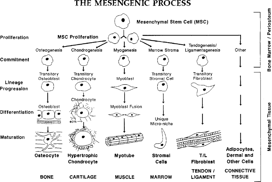
Adult marrow-derived mesenchymal stem cells (MSCs) are able to differentiate into bone, cartilage, muscle, marrow stroma, tendon-ligament, fat and other connective tissues. The questions can be asked, what do MSCs do naturally and where is the MSC niche? New insight and clinical experience suggest that MSCs are naturally found as perivascular cells, summarily referred to as pericytes, which are released at sites of injury, where they secrete large quantities of bioactive factors that are both immunomodulatory and trophic. The trophic activity inhibits ischaemia-caused apoptosis and scarring while stimulating angiogenesis and the mitosis of tissue intrinsic progenitor cells. The immunomodulation inhibits lymphocyte surveillance of the injured tissue, thus preventing autoimmunity, and allows allogeneic MSCs to be used in a variety of clinical situations. Thus, a new, enlightened era of experimentation and clinical trials has been initiated with xenogenic and allogeneic MSCs.
Conflict of interest statement
Conflict of interest statement: my colleagues and I started Osiris Therapeutics Inc. in late 1992. I no longer own stock in Osiris, neither do I have direct contact with the company.
Figures
References
-
- Caplan AI. Adult mesenchymal stem cells for tissue engineering versus regenerative medicine. J Cell Physiol 2007;213:341–347. - PubMed
-
- Caplan AI. Mesenchymal stem cells. J Orthop Res 1991;9:641–650. - PubMed
-
- Caplan AI. Mesenchymal stem cell: cell-based reconstructive therapy in orthopaedics. TissEng 2005;11:1198–1211. - PubMed
-
- Ed Thomas, Blume KG. Historical markers in the development of allogeneic hematopoietic cell transplantation. Biol Blood Marrow Transpl 1999;5:341–346. - PubMed
-
- Weiss L. The hematopoietic microenvironment of the bone marrow: an ultrastructural study of the stroma in rats. Anat Rec 1976;186:161–184. - PubMed
Publication types
MeSH terms
Grants and funding
LinkOut - more resources
Full Text Sources
Other Literature Sources
Medical
