

Short-term rescue of neonatal lethality in a mouse model of propionic acidemia by gene therapy
- PMID: 19025475
- PMCID: PMC2922073
- DOI: 10.1089/hum.2008.158
Short-term rescue of neonatal lethality in a mouse model of propionic acidemia by gene therapy
Abstract
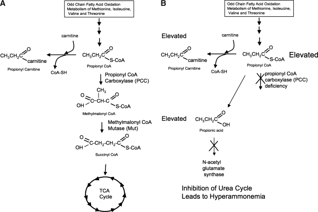
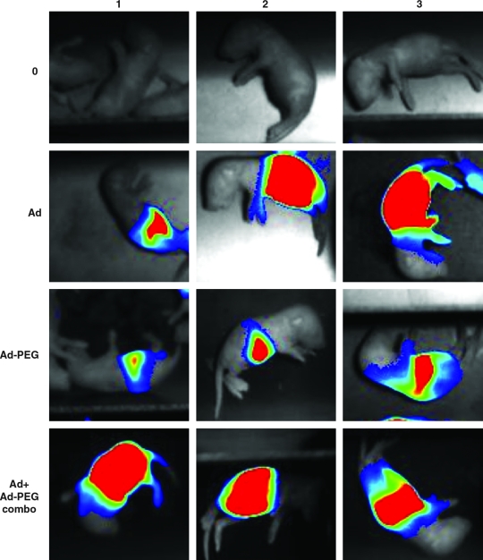
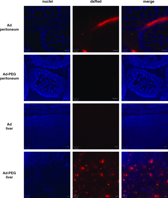
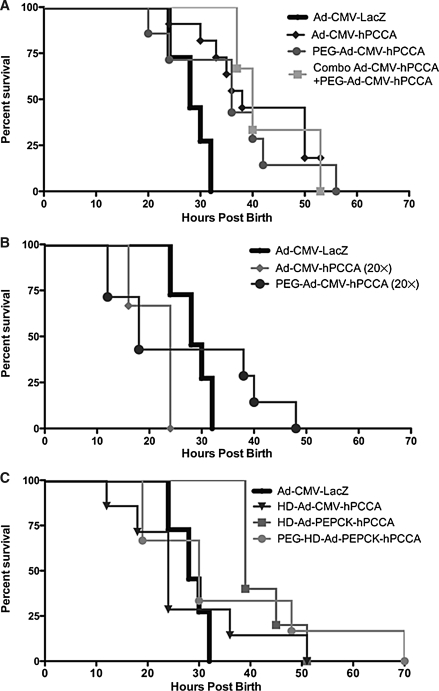
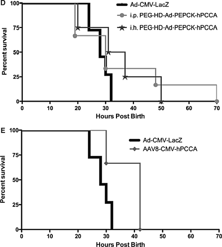
Propionic acidemia (PA) is a metabolic disorder that causes mental retardation and that can be fatal if untreated. PA is inherited in an autosomal recessive fashion involving mutations in PCCA or PCCB encoding the alpha and beta subunits of propionyl-CoA carboxylase (PCC). Current treatment is based on dietary restriction of substrate amino acids, which attenuates symptoms. However, patients still experience episodes of hyperammonemia that can cause progressive neurologic damage. In this paper, we have tested gene therapy approaches to PA in a stringent mouse model of PCCA deficiency, in which homozygous knockout mice are born but die within 36 hr. In this work, we have delivered first-generation and helper-dependent adenovirus serotype 5 (Ad5) vectors expressing the human PCCA cDNA by intraperitoneal injection into newborn mice. Unmodified Ad5 vectors mediated extensive transduction of the peritoneum with weak liver transduction as determined by luciferase imaging and dsRed expression. In contrast, modification of Ad5 with polyethylene glycol detargeted the virus from the peritoneum and retargeted it for transduction in the liver. When vectors expressing PCCA were injected, significant increases in life span were observed for both the unmodified and polyethylene glycol (PEG)-modified Ad5 vectors. However, this rescue was transient. Similarly, adeno-associated virus serotype 8-mediated transduction also produced only transient rescue. These data show first proof of principle for gene therapy of PA and demonstrate the potential utility of PEG to modify viral tropism in an actual gene therapy application.
Figures
Similar articles
-
Adeno-associated virus serotype 8 gene transfer rescues a neonatal lethal murine model of propionic acidemia.Hum Gene Ther. 2011 Apr;22(4):477-81. doi: 10.1089/hum.2010.164. Epub 2011 Feb 16. Hum Gene Ther. 2011. PMID: 20950151 Free PMC article.
-
Effects of adeno-associated virus serotype and tissue-specific expression on circulating biomarkers of propionic acidemia.Hum Gene Ther. 2014 Sep;25(9):837-43. doi: 10.1089/hum.2014.012. Epub 2014 Aug 21. Hum Gene Ther. 2014. PMID: 25046265 Free PMC article.
-
Generation of a hypomorphic model of propionic acidemia amenable to gene therapy testing.Mol Ther. 2013 Jul;21(7):1316-23. doi: 10.1038/mt.2013.68. Epub 2013 May 7. Mol Ther. 2013. PMID: 23648696 Free PMC article.
-
Propionic acidemia in the Arab World.Gene. 2015 Jun 15;564(2):119-24. doi: 10.1016/j.gene.2015.04.019. Epub 2015 Apr 9. Gene. 2015. PMID: 25865301 Review.
-
Pathophysiological mechanisms of complications associated with propionic acidemia.Pharmacol Ther. 2023 Sep;249:108501. doi: 10.1016/j.pharmthera.2023.108501. Epub 2023 Jul 22. Pharmacol Ther. 2023. PMID: 37482098 Free PMC article. Review.
Cited by
-
Helper-dependent adenoviral vectors for liver-directed gene therapy.Hum Mol Genet. 2011 Apr 15;20(R1):R7-13. doi: 10.1093/hmg/ddr143. Epub 2011 Apr 5. Hum Mol Genet. 2011. PMID: 21470977 Free PMC article. Review.
-
Relief of CoA sequestration and restoration of mitochondrial function in a mouse model of propionic acidemia.J Inherit Metab Dis. 2023 Jan;46(1):28-42. doi: 10.1002/jimd.12570. Epub 2022 Nov 3. J Inherit Metab Dis. 2023. PMID: 36251252 Free PMC article.
-
Dysregulated miRNAs and their pathogenic implications for the neurometabolic disease propionic acidemia.Sci Rep. 2017 Jul 18;7(1):5727. doi: 10.1038/s41598-017-06420-8. Sci Rep. 2017. PMID: 28720782 Free PMC article.
-
Metabolic flux analysis in hiPSC-CMs reveals insights into cardiac dysfunction in propionic acidemia Eva Richard.Res Sq [Preprint]. 2025 Jan 28:rs.3.rs-5874705. doi: 10.21203/rs.3.rs-5874705/v1. Res Sq. 2025. Update in: Cell Mol Life Sci. 2025 Apr 02;82(1):137. doi: 10.1007/s00018-025-05661-5. PMID: 39975893 Free PMC article. Updated. Preprint.
-
Perinatal gene transfer to the liver.Curr Pharm Des. 2011;17(24):2528-41. doi: 10.2174/138161211797247541. Curr Pharm Des. 2011. PMID: 21774770 Free PMC article. Review.
References
-
- Akiyama M. Thorne S. Kirn D. Roelvink P.W. Einfeld D.A. King C.R. Wickham T.J. Ablating CAR and integrin binding in adenovirus vectors reduces nontarget organ transduction and permits sustained bloodstream persistence following intraperitoneal administration. Mol. Ther. 2004;9:218–230. - PubMed
-
- Al Essa M. Rahbeeni Z. Jumaah S. Joshi S. Al Jishi E. Rashed M.S. Al Amoudi M. Ozand P.T. Infectious complications of propionic acidemia in Saudi Arabia. Clin. Genet. 1998;54:90–94. - PubMed
-
- Barshes N.R. Vanatta J.M. Patel A.J. Carter B.A. O'Mahony C.A. Karpen S.J. Goss J.A. Evaluation and management of patients with propionic acidemia undergoing liver transplantation: A comprehensive review. Pediatr. Transplant. 2006;10:773–781. - PubMed
-
- Campeau E. Desviat L.R. Leclerc D. Wu X. Perez B. Ugarte M. Gravel R.A. Structure of the PCCA gene and distribution of mutations causing propionic acidemia. Mol. Genet. Metab. 2001;74:238–247. - PubMed
Publication types
MeSH terms
Substances
LinkOut - more resources
Full Text Sources
Medical
