

Outward bulging of the right parietal bone in connection with fibrous dysplasia in an infant: a case report
- PMID: 19025620
- PMCID: PMC2611982
- DOI: 10.1186/1757-1626-1-347
Outward bulging of the right parietal bone in connection with fibrous dysplasia in an infant: a case report
Abstract
Background: Fibrous dysplasia (FD) is a developmental disease of bone in which there is replacement of normal spongiosa and filling of the medullary cavity of affected bones by an abnormal fibrous tissue that contains trabeculae of poorly calcified primitive bone formed by osseous metaplasia. Fibrous dysplasia is a common benign bone disease existing in monostotic and polyostotic forms. It is sometimes associated with aneurysmal bone cysts, and it is a component of McCune-Albright and Mazabraud syndromes.
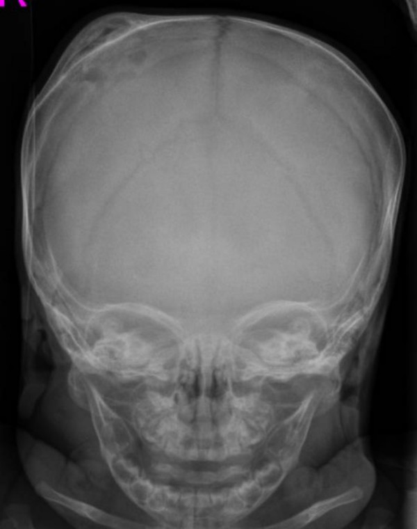
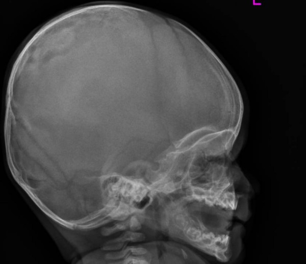
Case presentation: We describe here a 4-months old Austrian infant who presented with a hard bulging painless mass of (5 x 3 cm) of the right parietal bone. Radiographs showed a large irregular osteolytic lesion. T1-weighted MR image showed significant expansile lesion associated with a dense zone of calcification in the diploic space. To the best of our knowledge this is the first clinical report of an infant with early presentation of monostotic fibrous dysplasia of the right parietal bone.
Conclusion: Fibrous dysplasia of the skull is a painless progressively expanding destructive bone swellings produce cosmetic deformities. The clinical course may be unpredictable, with sudden appearance of symptoms, some of which can be important and irreversible. In our present patient, the possibility that an early surgical correction might positively interfere with the natural history of the lesion has to be evaluated by taking into account the obvious difficulties that will be encountered in reconstructing the skull after a wide excision of the pathologic bone.
Figures


References
-
- Lichtenstein L. Polyostotic fibrous dysplasia. Arch Surg. pp. 874–898.
-
- McCune D, Bruch H. Osteodystrophia fibrosa: report of a case in which the condition was combined with precocious puberty, multiple pigmentation of the skin and hyperthyroidism. AJDC. 1937;52:745–748.
-
- Faivre L, Nivelon-Chevalier A, Kottler ML, Robinet C, Khau van Kien P, Lorcerie B, Munnich A, Maroteaux P, Cormier-Daire V, Le Merrer M. Mazabraud syndrome in two patients: clinical overlap with McCune-Albright syndrome. Am J Med Genet. 2001;99:132–136. doi: 10.1002/1096-8628(2000)9999:999<00::AID-AJMG1135>3.0.CO;2-A. - DOI - PubMed
-
- Jee WH, Choi KH, Choe BY, Park JM, Shinn KS. Fibrous dysplasia: MR imaging characteristics with radiopathologic correlation. AJR Am J Roentgenol. 1996;167:1523–7. - PubMed
LinkOut - more resources
Full Text Sources
