. 2008 Nov 24:9:69.
doi: 10.1186/1471-2172-9-69.

Tolerance induced via TLR2 and TLR4 in human dendritic cells: role of IRAK-1

Affiliations

Tolerance induced via TLR2 and TLR4 in human dendritic cells: role of IRAK-1

Valerie Albrecht et al. BMC Immunol. .

Abstract

Background: While dendritic cells (DCs) can induce tolerance in T cells, little is known about tolerance induction in DCs themselves. We have analysed tolerance induced in human in-vitro generated DCs by repeated stimulation with ligands for TLR4 and TLR2.

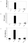
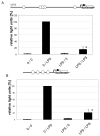
Results: DCs stimulated with the TLR4 ligand LPS did show a rapid and pronounced expression of TNF mRNA and protein. When DCs were pre-cultured for 2 days with 5 ng LPS/ml then the subsequent response to stimulation with a high dose of LPS (500 ng/ml) was strongly reduced for both TNF mRNA and protein. At the promoter level there was a reduced transactivation by the -1173 bp TNF promoter and by a construct with a tetrameric NF-kappaB motif. Within the signalling cascade leading to NF-kappaB activation we found an ablation of the IRAK-1 adaptor protein in LPS-tolerant DCs. Pre-culture of DCs with the TLR2 ligand Pam3Cys also led to tolerance with respect to TNF gene expression and IRAK-1 protein was ablated in such tolerant cells as well, while IRAK-4 protein levels were unchanged.

Conclusion: These data show that TLR-ligands can render DCs tolerant with respect to TNF gene expression by a mechanism that likely involves blockade of signal transduction at the level of IRAK-1.


Figures

Figure 1
Figure 1
Cell surface phenotype of monocyte-derived dendritic cells. Adherent mononuclear cells were cultured for 7 days with GM-CSF and IL-13 with addition of PGE2 for the final 2 days. Cells were then stained with specific monoclonal antibodies or the respective isotype controls and analysed in flow cytometry. Percentage of positive cells for CD1a were 48.4 ± 20.7%, for CD83 39.9 ± 14.5% and for CD209 89.5 ± 8.1%. Shown is one representative of 9 experiments.
Figure 2
Figure 2
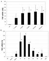
Dose response and time course analysis for LPS induced TNF mRNA in monocyte-derived DCs. A. Day 7 dendritic cells were stimulated with different doses of LPS from Salmonella abortus equi and after 2 hours TNF mRNA levels were determined by RT-PCR. All mRNA levels were adjusted to α- Enolase as house keeping gene. TNF mRNA expression was then calculated in AU while setting the value for TNF levels in unstimulated cells as one. Given is mean of 3 experiments. * = p < 0.05 compared to w/o. B. Dendritic cells were stimulated for different times with 500 ng/ml of LPS from Salmonella abortus equi and TNF mRNA levels were determined by RT-PCR. The maximum TNF mRNA expression level at 2 hours was set as 100%. Adjustment to α- Enolase was done as mentioned for Fig. 2A. Given is mean of 3 experiments. * = p < 0.05 compared to 0 h.
Figure 3
Figure 3
Induction of LPS tolerance in dendritic cells.A. Induction of LPS tolerance in dendritic cell cultures at the level of TNF mRNA at day 7 dendritic cells were cultured for additional 2 days with LPS at 5 ng/ml and then washed and stimulated at 500 ng LPS/ml. Cells were lysed at 2 hours and RNA was extracted, transcribed into cDNA and amplified by RT-PCR and data were normalized relative to levels of the house keeping gene alpha-Enolase. Given is the average of 7 experiments. * = p < 0.05 compared to 0/LPS. B. Induction of LPS tolerance in dendritic cell cultures at the level of TNF protein. Day 7 dendritic cells were cultured for additional 2 days with LPS at 5 ng/ml and then washed and stimulated at 500 ng LPS/ml for 6 hours. Supernatants were then harvested and were tested for TNF protein by ELISA. Given is the average of 3 experiments. * = p < 0.05 compared to 0/LPS. C. Induction of LPS tolerance in CD83+ dendritic cells at the level of TNF mRNA. Day 7 CD83+ dendritic cells were isolated by MACS to > 90% purity and these cells were then cultured for additional 2 days with LPS at 5 ng/ml, washed and stimulated at 500 ng LPS/ml. Cells were lysed and RNA was extracted, transcribed into cDNA and amplified by RT-PCR and data were normalized relative to levels of the house keeping gene alpha-Enolase. Given is the average of 3 experiments. * = p < 0.05 compared to 0/LPS.
Figure 4
Figure 4
A. Reduced TNF promoter activity in LPS tolerant dendritic cells. Day 7 dendritic cells were pre-cultured without and with LPS at 5 ng/ml and cells were infected on day 8 with the human – 1173 bp TNF promoter luciferase reporter adenovirus. On day 9 cells were stimulated for 5 hours with LPS at 500 ng/ml and luciferase activity was determined and normalized to protein content. Given is the average of 4 experiments. 100% represents 329560 +/- 373169 RLU in these experiments. * = p < 0.05 compared to 0/LPS. The hexagones denote NF-κB binding sites in the human TNF promoter at -873, a complex site from -627 to -589 including a site at – 605, and a site at -104 relative to transcription start. B. Reduced NF-κB activity in LPS tolerant dendritic cells. Day 7 dendritic cells were pre-cultured without and with LPS at 5 ng/ml and cells were infected on day 8 with a luciferase reporter adenovirus driven by a tetramer of NF-κB motifs. On day 9 cells were stimulated for 5 hours with LPS at 500 ng/ml and luciferase activity was determined and normalized to protein content. Given is the average of 3 experiments. 100% represents 13803,67 +/- 2651,93 RLU in these experiments. * = p < 0.05 compared to 0/LPS. The hexagones denote NF-κB binding sites.
Figure 5
Figure 5
Effect of LPS tolerance induction on levels of IRAK1 protein. Day 7 dendritic cells were cultured for additional 2 days with LPS at 5 ng/ml and then washed and stimulated at 500 ng LPS/ml. Lysates were prepared at 1 h and were subjected to Western blotting for IRAK-1 (A) or SHIP (B). One representative of 4 experiments.
Figure 6
Figure 6
Induction of LPS tolerance in dendritic cell cultures: effect on IL-12 mRNA. Day 7 dendritic cells were cultured for additional 2 days with LPS at 5 ng/ml and then washed and stimulated at 500 ng LPS/ml. Cells were lysed at 6 hours and RNA was extracted, transcribed into cDNA and amplified by RT-PCR and data were normalized relative to levels of the house keeping gene alpha-Enolase. Given is the average of 3 experiments. * = p < 0.05 compared to 0/LPS.
Figure 7
Figure 7
Induction of Pam3Cys tolerance in dendritic cells and effect on levels of IRAK1 protein. Day 7 dendritic cells were cultured for additional 2 days with Pam3Cys at 1μg/ml and then washed and stimulated at 10 μg Pam3Cys/ml. A. Cells were lysed at 2 hours and RNA was extracted, transcribed into cDNA and amplified by RT-PCR and data were normalized relative to levels of the house keeping gene alpha-Enolase. Given is the average of 5 experiments. * = p < 0.05 compared to 0/LPS. B. Lysates were prepared at 1 h and were subjected to Western blotting for IRAK-1. One representative of 3 experiments. C. Lysates were prepared at 1 h and were subjected to Western blotting for IRAK-4. One representative of 3 experiments.

References

    1. Ziegler-Heitbrock HW, Wedel A, Schraut W, Strobel M, Wendelgass P, Sternsdorf T, Bauerle PA, Haas JG, Riethmuller G. Tolerance to lipopolysaccharide involves mobilization of nuclear factor kappa B with predominance of p50 homodimers. J Biol Chem. 1994;269:17001–4. - PubMed
    1. Kastenbauer S, Ziegler-Heitbrock HW. NF-kappaB1 (p50) is upregulated in lipopolysaccharide tolerance and can block tumor necrosis factor gene expression. Infect Immun. 1999;67:1553–9. - PMC - PubMed
    1. Adib-Conquy M, Cavaillon JM. Gamma interferon and granulocyte/monocyte colony-stimulating factor prevent endotoxin tolerance in human monocytes by promoting interleukin-1 receptor-associated kinase expression and its association to MyD88 and not by modulating TLR4 expression. J Biol Chem. 2002;277:27927–34. doi: 10.1074/jbc.M200705200. - DOI - PubMed
    1. Li L, Cousart S, Hu J, McCall CE. Characterization of interleukin-1 receptor-associated kinase in normal and endotoxin-tolerant cells. J Biol Chem. 2000;275:23340–5. doi: 10.1074/jbc.M001950200. - DOI - PubMed
    1. Siedlar M, Frankenberger M, Benkhart E, Espevik T, Quirling M, Brand K, Zembala M, Ziegler-Heitbrock L. Tolerance induced by the lipopeptide Pam3Cys is due to ablation of IL-1R-associated kinase-1. J Immunol. 2004;173:2736–45. - PubMed

Publication types

MeSH terms

LinkOut - more resources