

Improved 3D phase contrast MRI with off-resonance corrected dual echo VIPR
- PMID: 19025882
- PMCID: PMC2778052
- DOI: 10.1002/mrm.21763
Improved 3D phase contrast MRI with off-resonance corrected dual echo VIPR
Abstract
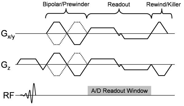
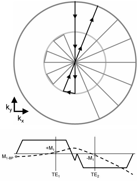
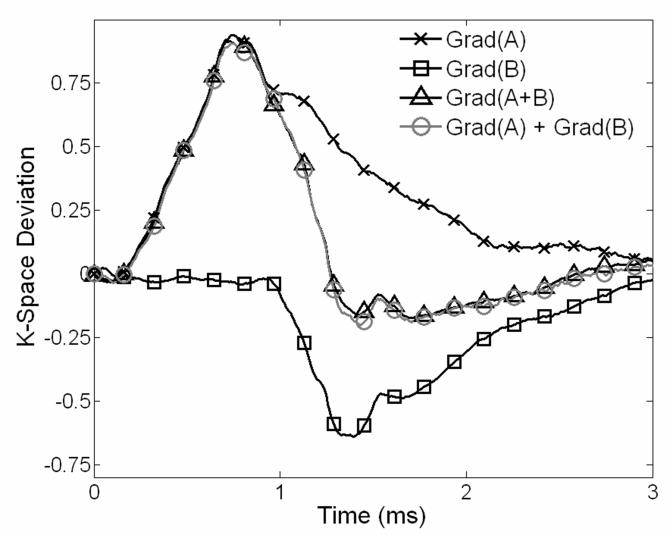
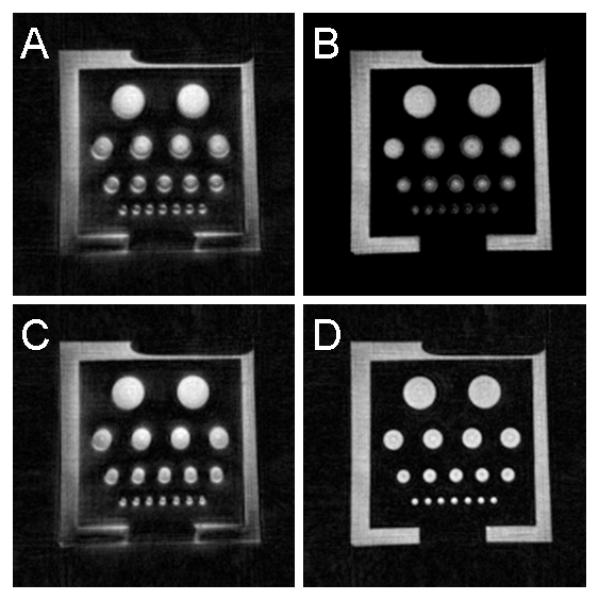
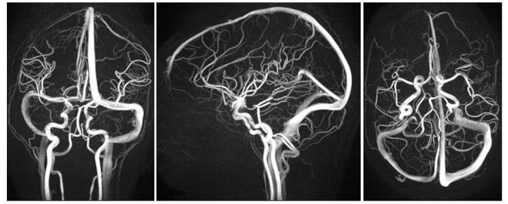
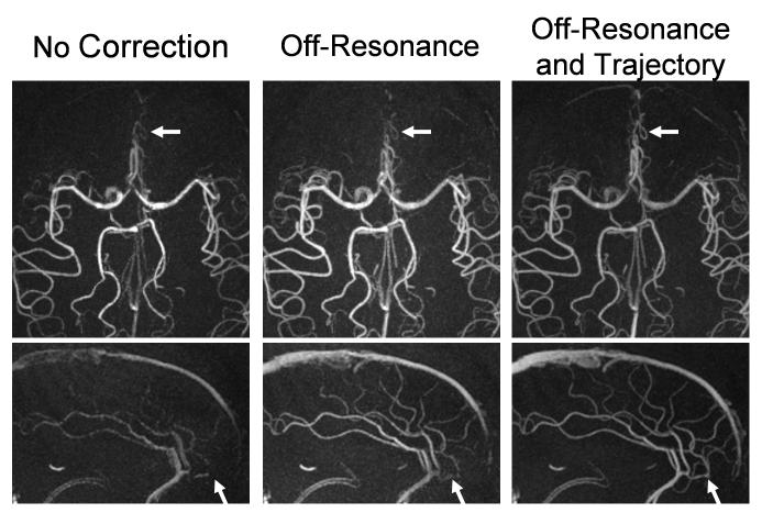
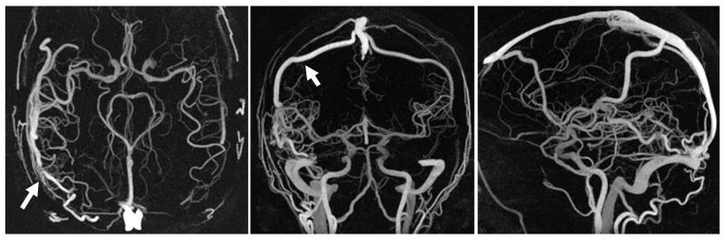
Phase contrast (PC) magnetic resonance imaging with a three-dimensional, radially undersampled acquisition allows for the acquisition of high resolution angiograms and velocimetry in dramatically reduced scan times. However, such an acquisition is sensitive to blurring and artifacts from off-resonance and trajectory errors. A dual-echo trajectory is proposed with a novel trajectory calibration from prescan data coupled with a multi-frequency reconstruction to correct for these errors. Comparisons of phantom data and in vivo results from volunteer, and patients with arteriovenous malformations patients are presented with and without these corrections and show significant improvement of image quality when both corrections are applied. The results demonstrate significantly improved visualization of vessels, allowing for highly accelerated PC acquisitions without sacrifice in image quality.
(c) 2008 Wiley-Liss, Inc.
Figures
References
-
- Huston J, Rufenacht DA, Ehman RL, Wiebers DO. Imtracranial Aneurysms and Vascular Malformations: Comparison of Time-of-Flight and Phase-Contrast Angiography. Radiology. 1991;181(3):721–730. - PubMed
-
- Oelerich M, Lentschig MG, Zunker P, Reimer P, Rummeney EJ, Schuierer G. Intracranial vascular stenosis and occlusion: comparison of 3D time-of-flight and 3D phase-contrast MR angiography. Nueroradiology. 1998;40(9):567–573. - PubMed
-
- Markl M, Draney MT, Miller DC, Levin JM, Williamson EE, Pelc NJ, Liang DH, Herfkens RJ. Time-resolved three-dimensional magnetic resonance velocity mapping of aortic flow in healthy volunteers and patients after valve-sparing aortic root replacement. J Thorac Cardiovasc Surg. 2005;130(2):456–463. - PubMed
-
- Tyszka JM, Laidlaw DH, Asa JW, Silverman JM. Three-dimensional, time-resolved (4D) relative pressure mapping using magnetic resonance imaging. J Magn Reson Imaging. 2000;12(2):321–329. - PubMed
-
- Papathanasopoulou P, Zhao S, Kohler U, Robertson MB, Long Q, Hoskins P, Xu XY, Marshall I. MRI measurement of time-resolved wall shear stress vectors in a carotid bifurcation model, and comparison with CFD predictions. J Magn Reson Imaging. 2002;17(2):153–162. - PubMed
Publication types
MeSH terms
Grants and funding
LinkOut - more resources
Full Text Sources
Other Literature Sources
Medical
