. 2008 Dec;83(6):744-51.
doi: 10.1016/j.ajhg.2008.11.001. Epub 2008 Nov 20.

Mutations in glucose transporter 9 gene SLC2A9 cause renal hypouricemia

Affiliations

Mutations in glucose transporter 9 gene SLC2A9 cause renal hypouricemia

Hirotaka Matsuo et al. Am J Hum Genet. 2008 Dec.

Erratum in

  • Am J Hum Genet. 2008 Dec;83(6):795

Abstract

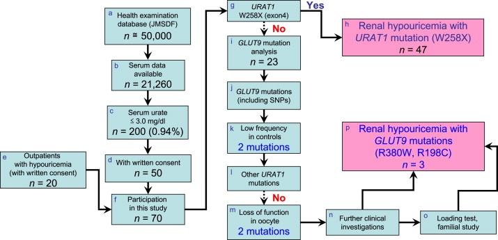
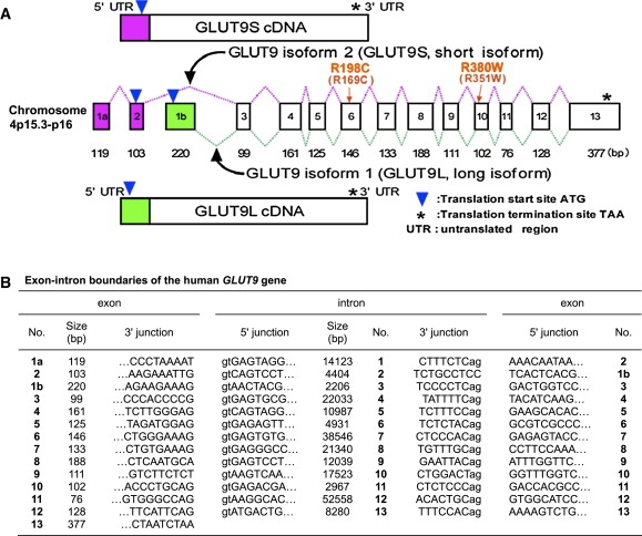
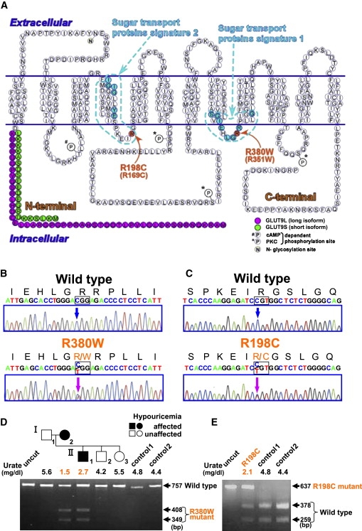
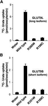
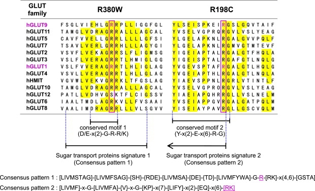
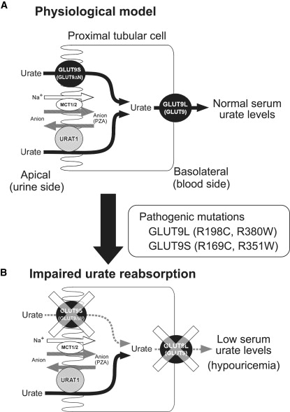
Renal hypouricemia is an inherited disorder characterized by impaired renal urate (uric acid) reabsorption and subsequent low serum urate levels, with severe complications such as exercise-induced acute renal failure and nephrolithiasis. We previously identified SLC22A12, also known as URAT1, as a causative gene of renal hypouricemia. However, hypouricemic patients without URAT1 mutations, as well as genome-wide association studies between urate and SLC2A9 (also called GLUT9), imply that GLUT9 could be another causative gene of renal hypouricemia. With a large human database, we identified two loss-of-function heterozygous mutations in GLUT9, which occur in the highly conserved "sugar transport proteins signatures 1/2." Both mutations result in loss of positive charges, one of which is reported to be an important membrane topology determinant. The oocyte expression study revealed that both GLUT9 isoforms showed high urate transport activities, whereas the mutated GLUT9 isoforms markedly reduced them. Our findings, together with previous reports on GLUT9 localization, suggest that these GLUT9 mutations cause renal hypouricemia by their decreased urate reabsorption on both sides of the renal proximal tubules. These findings also enable us to propose a physiological model of the renal urate reabsorption in which GLUT9 regulates serum urate levels in humans and can be a promising therapeutic target for gout and related cardiovascular diseases.


Figures

Figure 1
Figure 1
The Flowchart for Clinicogenetic Analysis of Hypouricemia with GLUT9 Mutations (a) We surveyed the health examination database of about 50,000 personnel of Japan Maritime Self-Defense Force (JMSDF). The database contains data for about 850,000 sets of examinations over 10 years (Apr. 1, 1997 to Mar. 31, 2007). To screen for renal hypouricemia, we selected 21,260 personnel data sets from 2006 in which serum urate data were available (b). (c) The number of persons who showed serum urate levels 3.0 mg/dl (178 μM) was 200 (0.94%) out of 21,260 persons. (d) 50 JMSDF persons who gave written consent and (e) an additional 20 outpatients with hypouricemia ([f] 70 hypouricemic cases in sum) participated in this clinicogenetic study. (g) First, we performed mutational analysis of URAT1 exon 4, to detect the most frequent mutation (W258X) in URAT1. (h) After 47 cases having the URAT1 W258X mutation were excluded, (i) remaining 23 hypouricemic cases were analyzed to find mutations in GLUT9. (j) After exclusion of known frequent SNPs or high-frequency mutations in Japanese controls, (k) 2 missense mutations (R380W and R198C) in GLUT9 were selected. (l) Subsequently, the patients with these GLUT9 mutations were confirmed to have no URAT1 mutations by whole-sequence analysis of URAT1. (m) Urate uptake activity was measured using oocytes, and these two mutations were proved to be loss-of-function mutations. (n) Further clinical investigations including a hypoxanthine assay and fractional excretion of urate (FEUA) were performed to confirm that the cases are true renal hypouricemia. (o) A pyrazinamide loading test and further familial studies were performed in possible cases. (p) Finally, two loss-of-function mutations in GLUT9 were identified as a cause of renal hypouricemia.
Figure 2
Figure 2
Genomic Structure of the Human GLUT9 Gene (A) The structure of the GLUT9 gene and cDNAs. The human GLUT9 gene contains 14 exons (1 noncoding and 13 coding) and is located on chromosome 4p15.3-p16. The alternative splicing of the GLUT9 gene results in two main transcripts: GLUT9 isoform 1 (long isoform, GLUT9L) and isoform 2 (short isoform, GLUT9S). (B) Exon-intron boundaries of the GLUT9 gene.
Figure 3
Figure 3
GLUT9 Mutations in Patients with Renal Hypouricemia (A) Mutation positions in a predicted human GLUT9 membrane topology model. Both mutations are present at equivalent positions within the cytoplasmic loops, which result in loss of positive charges. (B and C) Heterozygous mutations (c.1138C → T [p.R380W] and c.592C → T [p.R198C]; indicated by magenta arrows) were identified in the patients with renal hypouricemia. (D) The R380W mutation results in the gain of a BtsCI restriction site. A family tree of the hypouricemic patients having the R380W mutations is shown with serum urate levels. (E) The R198C mutation results in the loss of an AlwI site.
Figure 4
Figure 4
Markedly Reduced Urate Transport Activities in Oocytes that Express Mutant GLUT9 Isoforms High urate transport activities were observed in oocytes that express each wild-type GLUT9 isoform. In contrast, urate transport activity in oocytes was markedly reduced both in GLUT9L mutants (R380W and R198C) (A) and in GLUT9S mutants (R351W and R169C, which correspond to R380W and R198C in GLUT9L) (B). Results are expressed as mean ± SEM.
Figure 5
Figure 5
Amino Acid Conservation in the GLUT Family Transporters Arginine residues homologous to human GLUT9 amino acid positions 380 and 198, the sites of missense mutations identified in hypouricemia patients, are boxed in magenta. These mutations are observed at the well-known conserved motif (D/E-x(2)-G-R-R/K) and another conserved motif (Y-x(2)-E-x(6)-R-G) that is 100% conserved in all GLUT family transporters. These motifs are a part of the consensus patterns 1/2 that are demonstrated in the PROSITE database as “sugar transport proteins signatures 1/2” (also see Figure S3). The mutation sites in GLUT9 are found to be key residues in these consensus patterns. Interestingly, mutations of human GLUT1 at amino acid positions 333 and 153 (magenta), which correspond to the human GLUT9 mutation sites, are reported to cause GLUT1 deficiency syndrome. hHMIT represents human H+-coupled myo-inositol transporter, also known as SLC2A13.
Figure 6
Figure 6
Proposed Model of Renal Urate Reabsorption in Humans (A) Based on the findings from the hypouricemic patients with pathogenic GLUT9 mutations, we propose a physiological model of renal urate transport via human GLUT9 molecules. The localization of GLUT9L and GLUT9S is based on the previous observation from polarized MDCK cells. Here, GLUT9 mediates renal urate reabsorption on both sides of proximal tubular cells. URAT1 is expressed only on the apical side and is indirectly coupled with Na+-anion cotransporters, such as monocarboxylic acid transporter1/2 (MCT1/2). (B) An impaired urate reabsorption model in the renal proximal tubular cells. Pathogenic mutations in GLUT9L and GLUT9S on both sides of proximal tubules markedly reduce the urate reabsorption and cause hypouricemia. GLUT9L or “GLUT9” represents GLUT9 isoform 1 (long isoform) and the GLUT9S or GLUT9ΔN represents GLUT9 isoform 2 (short isoform). PZA represents pyrazinecarboxylic acid, a metabolite of pyrazinamide that is used for loading test of hypouricemic patients.

References

    1. Kikuchi Y., Koga H., Yasutomo Y., Kawabata Y., Shimizu E., Naruse M., Kiyama S., Nonoguchi H., Tomita K., Sasatomi Y. Patients with renal hypouricemia with exercise-induced acute renal failure and chronic renal dysfunction. Clin. Nephrol. 2000;53:467–472. - PubMed
    1. Diamond H.S., Paolino J.S. Evidence for a postsecretory reabsorptive site for uric acid in man. J. Clin. Invest. 1973;52:1491–1499. - PMC - PubMed
    1. Enomoto A., Kimura H., Chairoungdua A., Shigeta Y., Jutabha P., Cha S.H., Hosoyamada M., Takeda M., Sekine T., Igarashi T. Molecular identification of a renal urate anion exchanger that regulates blood urate levels. Nature. 2002;417:447–452. - PubMed
    1. Wakida N., Tuyen D.G., Adachi M., Miyoshi T., Nonoguchi H., Oka T., Ueda O., Tazawa M., Kurihara S., Yoneta Y. Mutations in human urate transporter 1 gene in presecretory reabsorption defect type of familial renal hypouricemia. J. Clin. Endocrinol. Metab. 2005;90:2169–2174. - PubMed
    1. Ichida K., Hosoyamada M., Hisatome I., Enomoto A., Hikita M., Endou H., Hosoya T. Clinical and molecular analysis of patients with renal hypouricemia in Japan-influence of URAT1 gene on urinary urate excretion. J. Am. Soc. Nephrol. 2004;15:164–173. - PubMed

Publication types

Substances

Associated data

LinkOut - more resources