. 2009 Mar;136(3):990-9.
doi: 10.1053/j.gastro.2008.10.047. Epub 2008 Oct 29.

Differentiation and transplantation of human embryonic stem cell-derived hepatocytes

Affiliations

Differentiation and transplantation of human embryonic stem cell-derived hepatocytes

Hesham Basma et al. Gastroenterology. 2009 Mar.

Abstract

Background & aims: The ability to obtain unlimited numbers of human hepatocytes would improve the development of cell-based therapies for liver diseases, facilitate the study of liver biology, and improve the early stages of drug discovery. Embryonic stem cells are pluripotent, potentially can differentiate into any cell type, and therefore could be developed as a source of human hepatocytes.

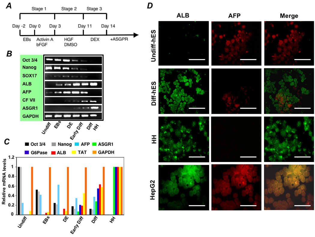
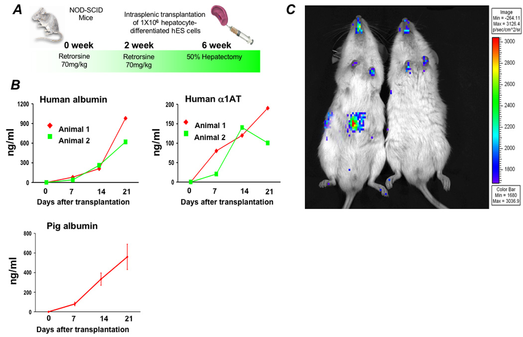
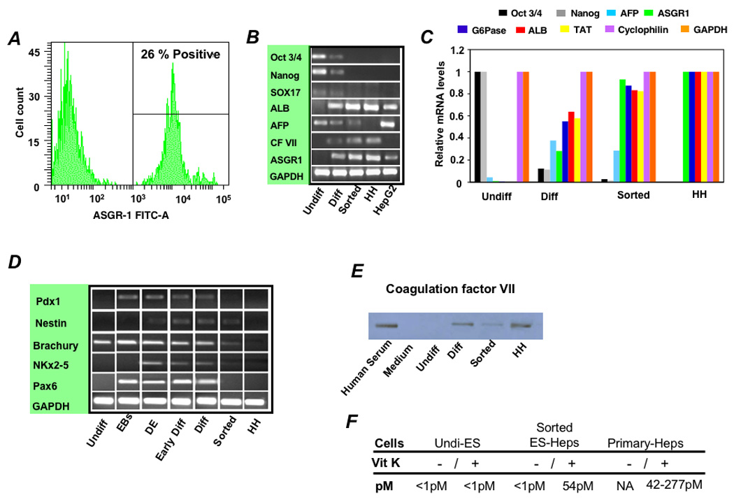
Methods: To generate human hepatocytes, human embryonic stem cells were differentiated by sequential culture in fibroblast growth factor 2 and human activin-A, hepatocyte growth factor, and dexamethasone. Functional hepatocytes were isolated by sorting for surface asialoglycoprotein-receptor expression. Characterization was performed by real-time polymerase chain reaction, immunohistochemistry, immunoblot, functional assays, and transplantation.

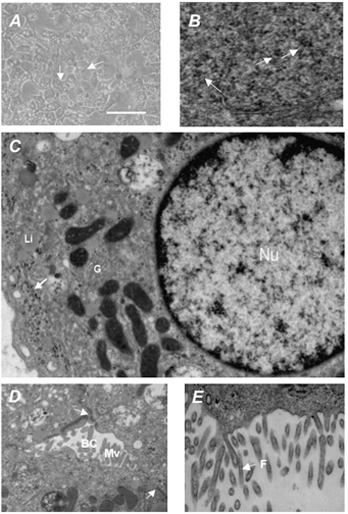
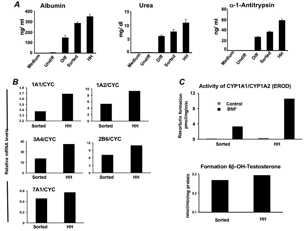
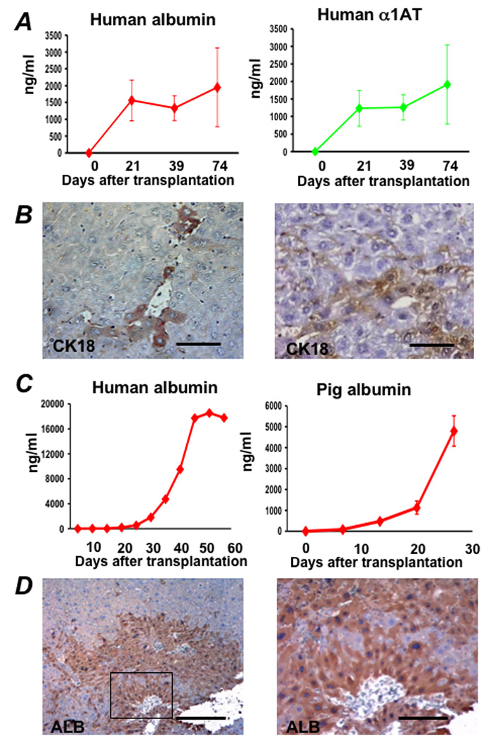
Results: Embryonic stem cell-derived hepatocytes expressed liver-specific genes, but not genes representing other lineages, secreted functional human liver-specific proteins similar to those of primary human hepatocytes, and showed human hepatocyte cytochrome P450 metabolic activity. Serum from rodents given injections of embryonic stem cell-derived hepatocytes contained significant amounts of human albumin and alpha1-antitrypsin. Colonies of cytokeratin-18 and human albumin-expressing cells were present in the livers of recipient animals.

Conclusions: Human embryonic stem cells can be differentiated into cells with many characteristics of primary human hepatocytes. Hepatocyte-like cells can be enriched and recovered based on asialoglycoprotein-receptor expression and potentially could be used in drug discovery research and developed as therapeutics.


Conflict of interest statement

Conflicts of interest: There are no conflicts of interest to disclose.

Figures

Figure 1
Figure 1. Differentiation of hES cells towards an hepatocytes phenotype and characterization of hepatocyte-directed cells
(A) Schematic representation of the strategy for differentiation of hES cells to hepatocytes. (B) RT-PCR and (C) real time analysis for expression of lineage-specific hepatic markers [albumin (ALB), ASGPR 1, glucose-6-phosphatase (G6Pase), tyrosine aminotransferase (TAT), and CF VII], endoderm markers (SOX 17 and AFP), and markers for undifferentiated cells (Pou5f1 and Nanog). Lanes: (1) Undifferentiated hES cells (Undiff), (2) following EB formation (EBs), (3) following treatment with FGF-2 and Activin-A or at the definitive endoderm (DE) stage, (4) following culture in HGF and DMSO (Early Diff), (5) and after culture in dexamethasone (Diff-ES). Primary human hepatocytes served as the positive control. Forward and reverse primers used for these studies are listed in the Supplementary Table 2. Immunohistochemistry of hES cells at day 18 of the differentiation protocol, after final exposure to dexamethasone, for albumin, AFP, and merged albumin and AFP expression (D). Undifferentiated hES (Undiff hES), primary human hepatocytes (HH), and HepG2 cells were used as controls. Scale bar 50 µm. Approximately 55% of cells expressed albumin while only 12% co-expressed AFP.
Figure 2
Figure 2. Morphological analysis of differentiated hES cells
Morphological analysis of differentiated hES cells in culture indicate that the differentiation program generated cells morphologically similar to hepatocytes, being polygonal in shape with multiple nuclei. (A) Transmission electron microscopy of differentiated hES showed (B) accumulation of glycogen rosettes (arrows), (C) round nuclei (Nu) with evenly distributed chromatin, Golgi complexes (G), and liposomes (Li), with glycogen rosettes identified again by an arrow, and (D, E) well developed bile canaliculi (BC) with apical microvilli (Mv) containing filaments (F) , tight junctions (arrows). Scale bar = 50 µm. Original magnification × 30,000.
Figure 3
Figure 3. Transplantation of differentiated cells and characterization of cells sorted for surface ASGPR expression
(A) One million hepatocyte-differentiated hES cells were transplanted into retrorsine-treated immune deficient NOD-SCID mice following 50% partial hepatectomy. Following transplantation, (B) human albumin and human α 1AT levels were measured by ELISA. Pig albumin levels in control mice transplanted with primary porcine hepatocytes are shown for comparison. (C) Imaging for luciferase expressing cells was performed three weeks after transplantation. Prior to transplantation, differentiated hES cells were transduced to express firefly luciferase. Differentiated cells containing an active albumin promoter, and thus expressing luciferase, appear to have traveled through the portal circulation after introduction into the spleen to engraft in the livers of transplanted mice. There was no evidence of luciferase-expressing cells in the spleen.
Figure 4
Figure 4. Enrichment of hES differentiated cells by ASGPR expression and characterization of sorted cells
(A) Flow cytometry showed that 18–26% of differentiated cells expressed the ASGPR, a definitive feature of differentiated hepatocytes. (B) RT-PCR and (C) Real-time analysis was performed to determine gene expression levels during the various stages of differentiation and following sorting relative to levels in primary human hepatocytes (HH). Expression levels of lineage-specific hepatic markers [albumin (ALB), tyrosine aminotransferase (TAT), glucose-6-phosphatase (G6P), CF VII, and ASGPR1], endoderm (Sox17) and the gestational hepatocyte marker AFP, and markers for undifferentiated cells (Pou5f1 and Nanog) were examined and mRNA expression levels were normalized relative to GAPDH, cyclophilin, and primary human hepatocytes. (D) To assess the lineage specificity of the differentiation program, mRNA was further analyzed by RT-PCR for Pdx-1 (a pancreatic marker gene), Nestin (ectoderm), Brachury (mesendoderm), NKx2.5 (mesoderm), and Pax-6 (ectoderm). Undifferentiated hES (lane 1), following EB formation (lane 2), hES differentiated to DE (lane 3), after culture in HGF and DMSO (early Diff; lane 4), after culture in dexamethasone (Diff; lane 5), and Diff stage cells following enrichment for ASGPR surface expression (sorted; lane 6). Primary human hepatocytes (lane 7) were used as control cells. (E) Production of CF VII protein by unsorted and ASGPR-sorted hES-derived cells was confirmed by immunoblot, and (F) functional CF VII activity in culture supernatants supplemented with Vitamin K was shown to be similar to that produced by cultured primary human hepatocytes.
Figure 5
Figure 5. Functional analyses of differentiated hES cells enriched toward a hepatocyte phenotype
(A) Albumin, urea, and alpha-1-antitrypsin secretion was determined in vitro by primary human hepatocytes (HH) and hES cells during differentiation (n= 5). Analysis involved undifferentiated hES cells (Undiff), hES cells following EB formation (EBs), hES cells after treatment with FGF-2 and Activin-A or at the definitive endoderm (DE) stage, hES cells after culture in HGF and DMSO (early Diff), hES cells after culture in dexamethasone (Diff), and hES cells after the Diff stage and following enrichment for ASGPR surface expression. (B) Real time analysis demonstrated expression of cytochrome P450 1A1 (CYP1A1), 1A2, 3A4, 2B6, and 7A1 at levels similar to that derived from fresh primary human hepatocytes. (C) To assess human liver-specific cytochrome P450 metabolic activity, ES-derived- or normal human hepatocytes were cultured in the presence of phenobarbital or 25 µM BNF. For measurement of CYP 1A activity, cells were exposed to media containing 20 µM 7-ethoxyresorufin and conversion to 7-hydroxyresorufin in the media was quantified by the fluorescence of the 7-hydroxy metabolite measured at 535 nm (Ex) and 581 nm (Em). Analysis of CYP3A activity was measured by conversion of testosterone to 6β-hydroxytestosterone by high pressure liquid chromatography. Studies demonstrated BNF-inducible EROD activity at approximately 25–30% of that generated by primary human hepatocytes, and baseline formation of testosterone by differentiated ASGPR-enriched cells near that produced by cultured primary human hepatocytes.
Figure 6
Figure 6. Transplantation of ASGPR-enriched hES-derived hepatocytes in uPA-SCID mice and an immune suppressed Nagase analbuminemic rat
One hundred thousand to two-hundred thousand hES-derived cells, sorted based on surface ASGPR expression, were transplanted in immune deficient Alb-uPA SCID mice. Following transplantation, (A) human albumin and human α1AT levels were measured by ELISA. At 75 days after transplantation, the serum of these animals contained 1000–2000 ng/ml human albumin and AAT. (B) Immunohistochemistry was performed on liver sections of transplanted animals, and small clusters of human cytokeratin-18 (CK18)-staining engrafted cells, which stain dark brown, with hepatocyte morphology were present throughout the liver. (C) One million ASGPR-sorted differentiated hES cells were also transplanted into the spleen of a retrorsine-treated FK506 immune suppressed Nagase analbuminemic rat that underwent 70% partial hepatectomy at the time of transplantation. Following transplantation, human albumin levels were measured at 20,000 ng/ml (0.2 mg/ml) by ELISA 55 days after transplantation. Comparable serum porcine albumin levels were measured in control Alb-uPA SCID mice up to 28 days after transplantation with 4°C 24-hour University of Wisconsin preserved primary pig hepatocytes. At sacrifice, immunohistochemistry demonstrated large colonies of human albumin-expressing cells (D).

Comment in

References

    1. Matas AJ, Sutherland DE, Steffes MW, Mauer SM, Sowe A, Simmons RL, Najarian JS. Hepatocellular transplantation for metabolic deficiencies: decrease of plasms bilirubin in Gunn rats. Science. 1976;192:892–894. - PubMed
    1. Groth CG, Arborgh B, Bjorken C, Sundberg B, Lundgren G. Correction of hyperbilirubinemia in the glucuronyltransferase-deficient rat by intraportal hepatocyte transplantation. Transplant Proc. 1977;9:313–316. - PubMed
    1. Fox IJ, Roy-Chowdhury J. Hepatocyte transplantation. J Hepatol. 2004;40:878–886. - PubMed
    1. Azuma H, Paulk N, Ranade A, Dorrell C, Al-Dhalimy M, Ellis E, Strom S, Kay MA, Finegold M, Grompe M. Robust expansion of human hepatocytes in Fah−/−/Rag2−/−/Il2rg−/− mice. Nat Biotechnol. 2007;25:903–910. - PMC - PubMed
    1. Mercer DF, Schiller DE, Elliott JF, Douglas DN, Hao C, Rinfret A, Addison WR, Fischer KP, Churchill TA, Lakey JR, Tyrrell DL, Kneteman NM. Hepatitis C virus replication in mice with chimeric human livers. Nat Med. 2001;7:927–933. - PubMed

Publication types

MeSH terms