. 2009 Mar;156(1-2):19-26.
doi: 10.1016/j.jviromet.2008.10.022. Epub 2008 Dec 5.

Human airway epithelial cell culture to identify new respiratory viruses: coronavirus NL63 as a model

Affiliations

Human airway epithelial cell culture to identify new respiratory viruses: coronavirus NL63 as a model

Bridget S Banach et al. J Virol Methods. 2009 Mar.

Abstract

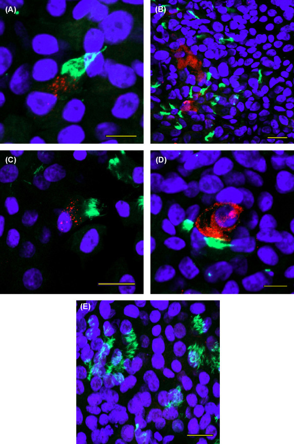
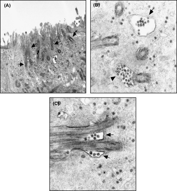
Propagation of new human respiratory virus pathogens in established cell lines is hampered by a lack of predictability regarding cell line permissivity and by availability of suitable antibody reagents to detect infection in cell lines that do not exhibit significant cytopathic effect. Recently, molecular methods have been used to amplify and identify novel nucleic acid sequences directly from clinical samples, but these methods may be hampered by the quantity of virus present in respiratory secretions at different time points following the onset of infection. Human airway epithelial (HAE) cultures, which effectively mimic the human bronchial environment, allow for cultivation of a wide variety of human respiratory viral pathogens. The goal of the experiments described here was to determine if propagation and identification of a human respiratory virus may be achieved through inoculation of HAE cultures followed by whole transcriptome amplification (WTA) and sequence analysis. To establish proof-of-principle human coronavirus NL63 (HCoV-NL63) was evaluated, and the first visualization of HCoV-NL63 virus by transmission electron microscopy (TEM) is reported. Initial propagation of human respiratory secretions onto HAE cultures followed by TEM and WTA of culture supernatant may be a useful approach for visualization and detection of new human respiratory pathogens that have eluded identification by traditional approaches.


Figures

Fig. 1
Fig. 1
Detection of HCoV-NL63 in the ciliated epithelium of HAE cultures by immunofluorescence. HCoV-NL63-infected (A–D) or mock-infected (E) HAE cultures were stained with anti-HCoV-NL63 replicase (red), anti-tubulin (green) and DAPI (blue) at 72 h post-infection and imaged by confocal microscopy. (A and D) Bar, 10 μm. (B, C and E) Bar, 20 μM.
Fig. 2
Fig. 2
Detection of HCoV-NL63 using TEM. (A) HCoV-NL63 replication and budding into vesicles occurs in ciliated cells. The cilia (arrows) are tangentially sectioned; ×39,000. (B) HCoV-NL63 virions in a clear Golgi vacuole (arrow). A second vacuole (arrowhead) is at the base of a cilium. Single virions in small vacuoles are detected in the background; ×126,000 (C) Extracellular virions surround the base of two cilia (arrows). Single intravacuolar virions are in the background; ×146,000.
Fig. 3
Fig. 3
Budding of HCoV-NL63 into smooth-walled vesicles. (A) A budding virion is seen in a vacuole (arrow). Two other virions (arrowheads) have visible spikes, characteristic of members of the Coronaviridae; ×245,000. (B) A vacuole with several budding virions (arrows) and free particles covered with spikes (arrowheads); ×175,000.
Fig. 4
Fig. 4
Evidence of lytic infection by HCoV-NL63. (A) Section of a necrotic cells on the surface of an HAE culture. One of the vacuoles contains virions (arrow); ×33,000. (B) An electron-dense apoptotic cell with vesicles containing virions (arrow) is present within the HAE cell layer; ×35,000. (C) Enlargement of (B), showing the cell contains vacuoles (e.g. arrows) with virions; ×184,000.
Fig. 5
Fig. 5
Analysis of products of WTA reaction products generated from RNA isolated from the supernatant of mock-infected and HCoV-NL63 inoculated HAE cultures. (A) Ethidium bromide stain of WTA products from mock-infected (lane 1) and HCoV-NL63-infected (lane 2) cells; (B) analysis of products generated by restriction enzyme digestion (EcoRI) of cloned WTA products separated by electrophoresis on a 1.5% agarose gel and visualized by staining with ethidium bromide. (C) Sequence of clone #56 identified by BLAST as 98% homologous to HCoV-NL63.

References

    1. Afonso C.L. Sequencing of avian influenza virus genomes following random amplification. BioTechniques. 2007;43:188. - PubMed
    1. Allander T., Tammi M.T., Eriksson M., Bjerkner A., Tiveljung-Lindell A., Andersson B. Cloning of a human parvovirus by molecular screening of respiratory tract samples. Proc. Natl. Acad. Sci. U.S.A. 2005;102:12891–12896. - PMC - PubMed
    1. Arden K.E., Nissen M.D., Sloots T.P., Mackay I.M. New human coronavirus, HCoV-NL63, associated with severe lower respiratory tract disease in Australia. J. Med. Virol. 2005;75:455–462. - PMC - PubMed
    1. Bastien N., Anderson K., Hart L., Van Caeseele P., Brandt K., Milley D., Hatchette T., Weiss E.C., Li Y. Human coronavirus NL63 infection in Canada. J. Infect. Dis. 2005;191:503–506. - PMC - PubMed
    1. Bozzola J.J., Russell L.D. second ed. Jones and Bartlett Publishers; Sandbury, Mass: 1998. Electron Microscopy: Principles and Techniques for Biologists.

Publication types

LinkOut - more resources