

Influence of polymer structure and biodegradation on DNA release from silk-elastinlike protein polymer hydrogels
- PMID: 19027056
- PMCID: PMC2680215
- DOI: 10.1016/j.ijpharm.2008.10.021
Influence of polymer structure and biodegradation on DNA release from silk-elastinlike protein polymer hydrogels
Abstract
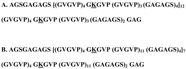
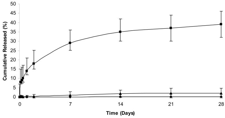
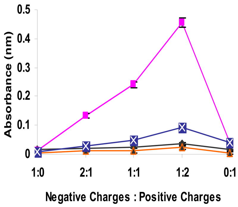
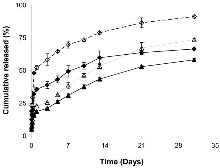
Silk-elastinlike protein polymers (SELPs) of varying ratios and lengths of silk and elastin blocks capable of hydrogel formation were evaluated as matrices for controlled delivery of plasmid DNA. Influence of polymer structure, ionic strength of the media and gelation time on DNA release from two structurally related hydrogels, SELP-47K and SELP-415K, was evaluated. The influence of elastase-induced degradation on the swelling behavior and DNA release from these hydrogels was investigated. Results indicate that release is a function of polymer structure, concentration and cure time. SELP-415K which has twice the number of elastin units as that of SELP-47K demonstrated higher release than that of SELP-47K. DNA release from these hydrogels is an inverse function of polymer concentration and cure time, with higher release observed at lower polymer concentration and shorter cure time. Results indicate that ionic strength of the media governs the rate of release. An increase in swelling ratio was observed in the presence of elastase at 12 wt.% composition for both SELP analogs. Release in the presence of elastase was enhanced due to increased swelling ratio and loss of hydrogel integrity. These studies allude to the utility of recombinant techniques to control plasmid DNA release and biodegradation in SELP hydrogels.
Figures
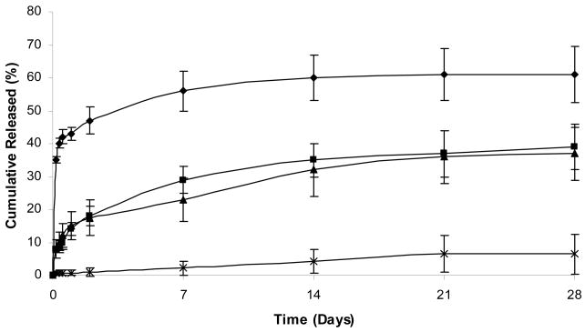
 ). Each symbol represents mean ± standard deviation for n=3 samples.
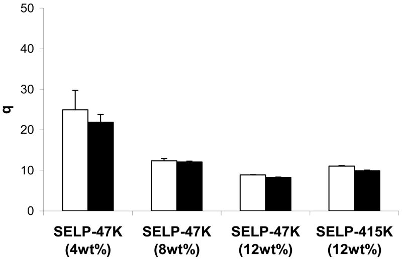
References
-
- Bellocq NC, Kang DW, Wang X, Jensen GS, Pun SH, Schluep T, Zepeda ML, Davis ME. Synthetic biocompatible cyclodextrin-based constructs for local gene delivery to improve cutaneous wound healing. Bioconjug Chem. 2004;15:1201–11. - PubMed
-
- Cappello J, Crissman J, Dorman M, Mikolajczak M, Textor G, Marquet M, Ferrari F. Genetic engineering of structural protein polymers. Biotechnol Prog. 1990;6:198–202. - PubMed
-
- Cohen-Sacks H, Elazar V, Gao J, Golomb A, Adwan H, Korchov N, Levy RJ, Berger MR, Golomb G. Delivery and expression of pDNA embedded in collagen matrices. J Control Release. 2004;95:309–320. - PubMed
-
- Dandu R, Cappello J, Ghandehari H. Characterization of structurally related adenovirus-laden silk-elastinlike hydrogels. J of Bioact and Comp Poly. 2008;23:5–19.
-
- Dandu R, Ghandehari H. Delivery of bioactive agents from recombinant polymers. Progress in Polymer Science. 2007;32:1008–1030.
Publication types
MeSH terms
Substances
Grants and funding
LinkOut - more resources
Full Text Sources
Other Literature Sources
Miscellaneous
