. 2009 Feb;136(2):551-63.
doi: 10.1053/j.gastro.2008.10.081. Epub 2008 Nov 6.

Targeted epithelial tight junction dysfunction causes immune activation and contributes to development of experimental colitis

Affiliations

Targeted epithelial tight junction dysfunction causes immune activation and contributes to development of experimental colitis

Liping Su et al. Gastroenterology. 2009 Feb.

Abstract

Background & aims: Inflammatory bowel disease (IBD) is a multifactorial disease thought to be caused by alterations in epithelial function, innate and adaptive immunity, and luminal microbiota. The specific role of epithelial barrier function remains undefined, although increased activity of intestinal epithelial myosin light chain kinase (MLCK), which is the primary mechanism of tumor necrosis factor-induced barrier dysfunction, occurs in human IBD. Our aim was to determine whether, in an intact epithelium, primary dysregulation of the intestinal epithelial barrier by pathophysiologically relevant mechanisms can contribute to development of colitis.

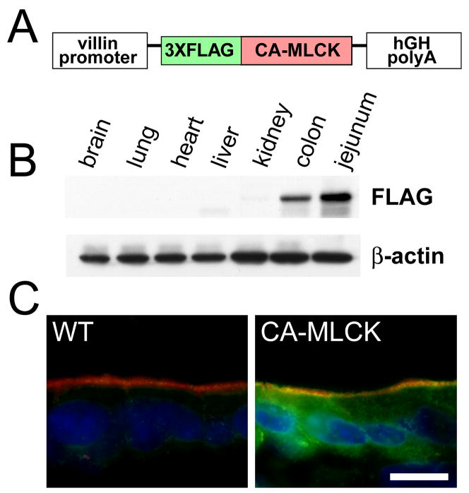
Methods: We developed transgenic (Tg) mice that express constitutively active MLCK (CA-MLCK) specifically within intestinal epithelia. Their physiology, immune status, and susceptibility to disease were assessed and compared with non-Tg littermate controls.

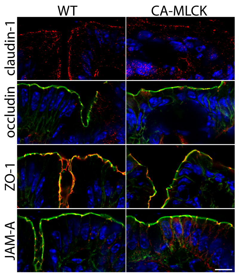
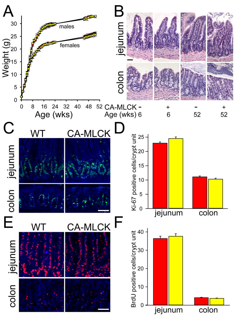
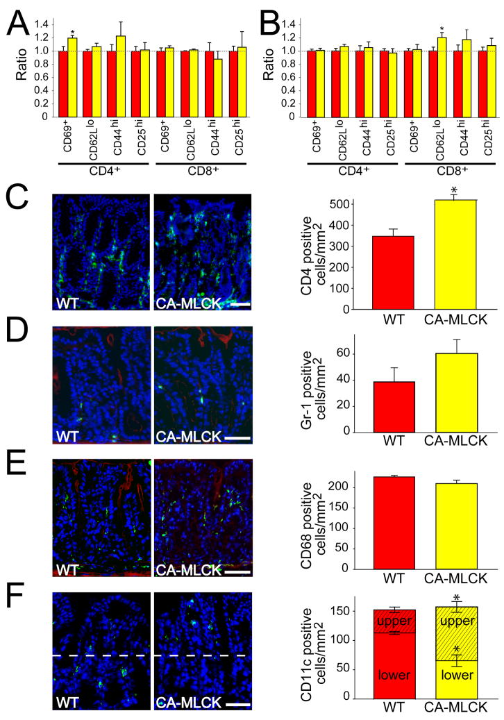
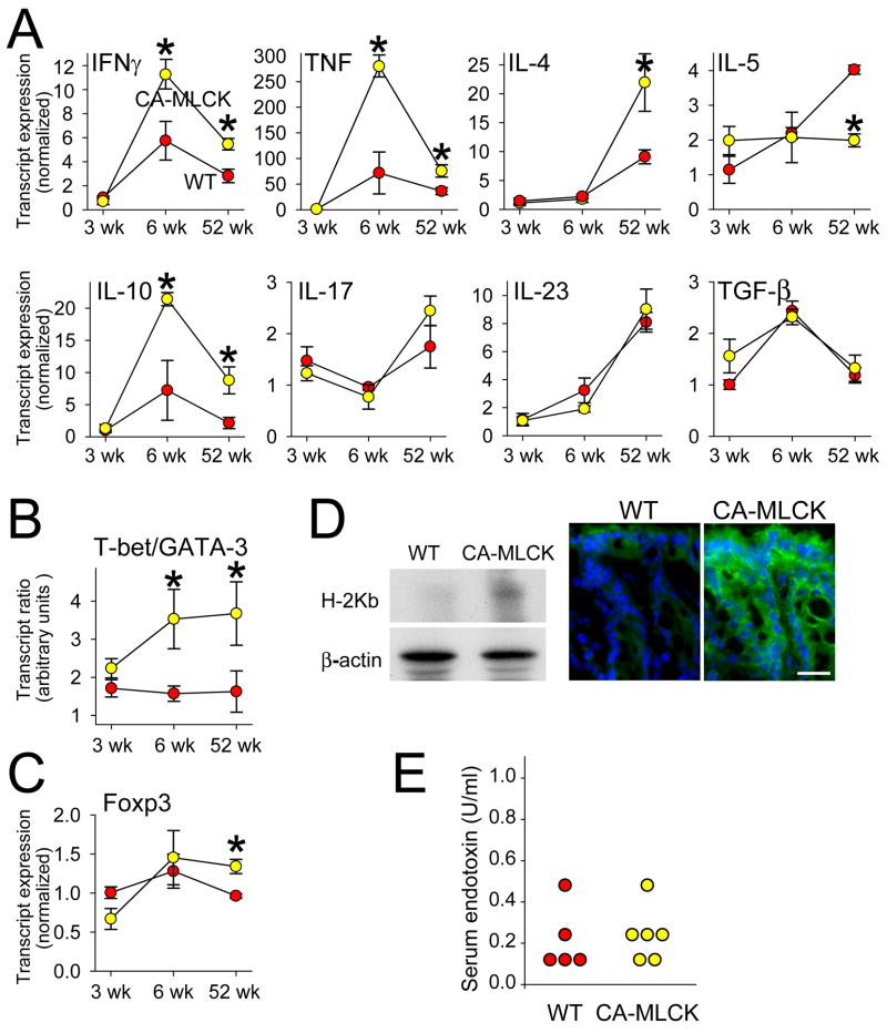
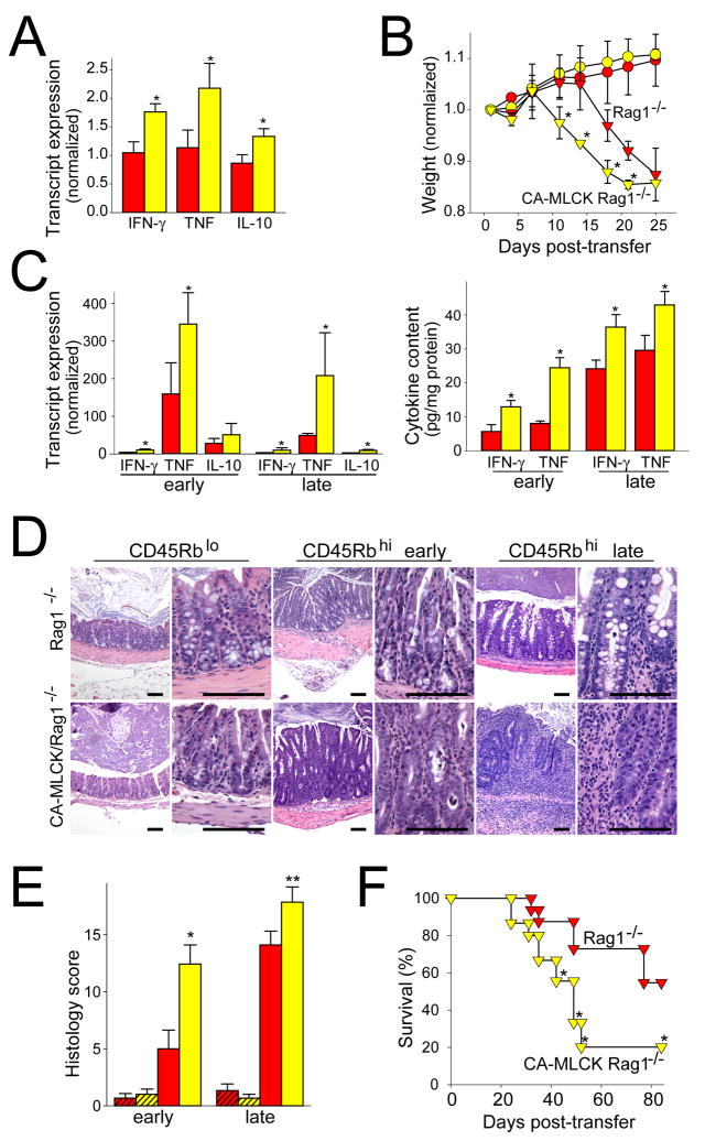
Results: CA-MLCK Tg mice demonstrated significant barrier loss but grew and gained weight normally and did not develop spontaneous disease. CA-MLCK Tg mice did, however, develop mucosal immune activation demonstrated by increased numbers of lamina propria CD4(+)lymphocytes, redistribution of CD11c+cells, increased production of interferon-gamma and tumor necrosis factor, as well as increased expression of epithelial major histocompatibility complex class I. When challenged with CD4+CD45+Rb(hi) lymphocytes, Tg mice developed an accelerated and more severe form of colitis and had shorter survival times than non-Tg littermates.

Conclusions: Primary pathophysiologically relevant intestinal epithelial barrier dysfunction is insufficient to cause experimental intestinal disease but can broadly activate mucosal immune responses and accelerate the onset and severity of immune-mediated colitis.


Conflict of interest statement

No conflicts of interest exist

Figures

Figure 1
Figure 1. Generation of CA-MLCK mice expressing constitutively-activate MLCK
(A) The CA-MLCK transgene construct. (B) Immunoblot of indicated organs from CA-MLCK Tg mice. (C) CA-MLCK, detected via the FLAG epitope tag (green), in jejunal epithelium. F-actin (red) and nuclei (blue) are shown. Bar, 10 μm.
Figure 2
Figure 2. CA-MLCK expression causes increas ed intestinal epithelial MLC phosphorylation and intestinal barrier loss
(A) Phosphorylated MLC (red) in jejunum and colon of WT and CA-MLCK Tg mice. Nuclei are shown in blue. Bar, 10 μm. (B, C) Immunoblot of phosphorylated MLC (pMLC) and total MLC and densitometric analysis of WT (red bars) and CA-MLCK Tg (yellow bars) after in vivo perfusion with (hatched bars) or without (solid bars) PIK. n=4 for each condition. *, P<0.01 vs. WT littermates; **, P<0.05 vs. CA-MLCK Tg mice without PIK. (D) Paracellular BSA flux with or without PIK. n=4 for each condition. *, P<0.01 vs. WT; **, P<0.01 vs. Tg without PIK.(E) In vivo measurement of colonic paracellular permeability in WT and CA-MLCK Tg mice. *, P<0.02 vs. WT.
Figure 3
Figure 3. CA-MLCK Tg mice do not display alterations in tight junction organization
Immunostains of tight junction-associated proteins in colonic mucosa of CA-MLCK Tg and WT mice. Junctional proteins (claudin-1, occludin, ZO-1, JAM-A) are shown in red. Nuclei are blue. F-actin is shown in green in the images of occludin, ZO-1, and JAM-A. Bar, 10 μm.
Figure 4
Figure 4. CA-MLCK Tg mice grow normally and do not display histological disease
(A) Growth of CA-MLCK Tg (yellow symbols) and WT (red symbols) mice (male, squares; female, circles). (B) Jejunum and proximal colon of CA-MLCK Tg and WT mice at 6 and 52 wks of age, respectively. Bar, 50 μm. (C, D) Ki-67 (green) in jejunum and proximal colon of CA-MLCK Tg (yellow bars) and WT (red bars) mice. Bar, 30 μm. (E, F) BrdU (red) incorporation and morphometry of CA-MLCK Tg (yellow bars) and WT (red bars) mice in jejunum and proximal colon epithelium after 36h and 2h, respectively. Bar, 30 μm.
Figure 5
Figure 5. Increased paracellular permeability modifies lamina propria immune cell recruitment
(A) Intraepithelial lymphocytes of WT (red bars) and CA-MLCK Tg (yellow bars) mice. *, P<0.02 vs. WT.(B) Lamina propria lymphocytes we re harvested from colon of WT (red bars) and CA-MLCK Tg (yellow bars) mice. *, P<0.02 vs. WT.(C) CD4 + cells (green) in the colonic lamina propria of WT and CA-MLCK Tg mice. Morphometric analysis of WT (red bar) and CA-MLCK Tg (yellow bar) mice is shown. Bar, 50 μm. *, P<0.03. (D) Gr-1+ cells (green) in the colonic lamina propria of WT and CA-MLCK Tg mice. F-actin is shown in red. Morphometric analysis is shown. Bar, 50 μm. (E) CD68+ cells (green) in the colonic lamina propria of WT and CA-MLCK Tg mice. F-actin is shown in red. Morphometric analysis is shown. Bar, 50 μm. (F) CD11c+ cells (green) in the colonic lamina propria of WT and CA-MLCK Tg mice. Morphometric analysis is shown right, separating superficial (hatched) and basal (solid) sections of the lamina propria. Bar, 40 μm. *, P<0.01.
Figure 6
Figure 6. Subclinical mucosal immune activation is present in CA-MLCK Tg mice
(A) Transcript content for indicated cytokines in proximal colon of WT (red symbols) and CA-MLCK Tg (yellow symbols) mice. *, P<0.03. (B) T-bet/GATA-3 transcript ratios in WT (red symbols) and CA-MLCK Tg (yellow symbols) mice at 3 wks, 6wks and 52 wks of age. *, P<0.02. (C) Foxp3 transcript expression in WT and Tg *, P<0.02. (D) H2Kb expression was assessed by SDS-PAGE and immunoblot in isolated colonic epithelial cells of WT and CA-MLCK mice. Immunofluorescence detection of H2Kb expression (green) in colonic epithelium of WT and CA-MLCK mice. Bar, 20 μm. (E) Endotoxin assay showed similar levels in serum of WT (red symbols) and CA-MLCK Tg (yellow symbols) mice.
Figure 7
Figure 7. Increased paracellular permeability accelerates immune-mediated colitis
(A) Transcript content in proximal colon of 6wk old RAG1−/− (red bars) and CA-MLCK Tg, RAG1−/− (yellow bars) mice, prior to adoptive transfer. *, P<0.01. (B) Weight after adoptive transfer of CD4+CD45Rbhi (triangles) and CD4+CD45Rblo (circles) T cells into RAG1−/− (red symbols) and CA-MLCK Tg, RAG1−/− (yellow symbols) recipients. Weights were normalized to weight prior to adoptive transfer. *, P<0.05 vs RAG1−/− recipients receiving CD4+CD45Rbhi T cells. (C) Transcript content in proximal colon of RAG1−/− (red bars) and CA-MLCK Tg, RAG1−/− (yellow bars) mice 12 d (early) or 19 d (late) after adoptive transfer. *, P<0.03. Changes in IFN-γ and TNF protein expression were similar. *, P<0.04 (D) Colon histology 12 d (early) and 19 d (late) after adoptive transfer. Bar, 100 μm. (E) Histology scores of the proximal colon sections of RAG1−/− (red bars) and CA-MLCK Tg, RAG1−/− (yellow bars) mice 12 d (early) and 19 d (late) after adoptive transfer. Mice received either CD4+CD45Rblo (hatched bars) or CD4+CD45Rbhi (solid bars) T cells. *, P<0.01 vs. RAG1−/− recipients transferred with CD4+CD45Rbhi T cells, ** P<0.03 vs. RAG1−/− recipients transferred with CD4+CD45Rbhi T cells. (F) Survival of RAG1−/− (red symbols) and CA-MLCK Tg, RAG1−/− (yellow symbols) recipients after transfer of CD4+CD45Rbhi T cells. *, P<0.05.

References

    1. Pearson AD, Eastham EJ, Laker MF, et al. Intestinal permeability in children with Crohn’s disease and coeliac disease. Br Med J (Clin Res Ed) 1982;285:20–1. - PMC - PubMed
    1. Hollander D, Vadheim CM, Brettholz E, et al. Increased intestinal permeability in patients with Crohn’s disease and their relatives. A possible etiologic factor. Ann Intern Med. 1986;105:883–5. - PubMed
    1. May GR, Sutherland LR, Meddings JB. Is small intestinal permeability really increased in relatives of patients with Crohn’s disease? Gastroenterology. 1993;104:1627–32. - PubMed
    1. Katz KD, Hollander D, Vadheim CM, et al. Intestinal permeability in patients with Crohn’s disease and their healthy relatives. Gastroenterology. 1989;97:927–31. - PubMed
    1. Clayburgh DR, Shen L, Turner JR. A porous defense: the leaky epithelial barrier in intestinal disease. Lab Invest. 2004;84:282–91. - PubMed

Publication types

MeSH terms

Substances