. 2009 Jan 30;385(4):1287-99.
doi: 10.1016/j.jmb.2008.11.002. Epub 2008 Nov 12.

The capsid proteins of a large, icosahedral dsDNA virus

Affiliations

The capsid proteins of a large, icosahedral dsDNA virus

Xiaodong Yan et al. J Mol Biol. .

Abstract

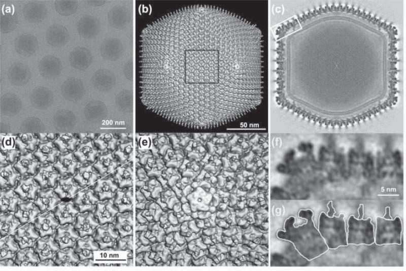
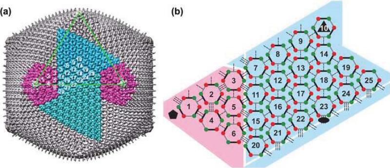
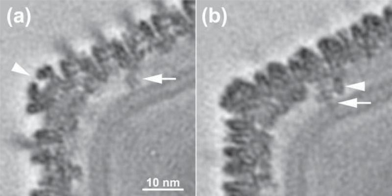
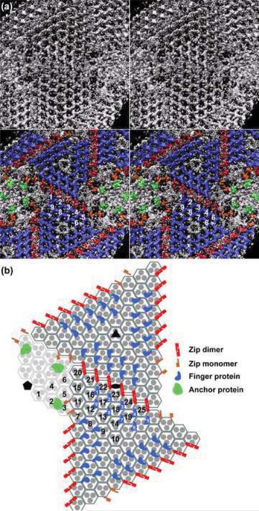
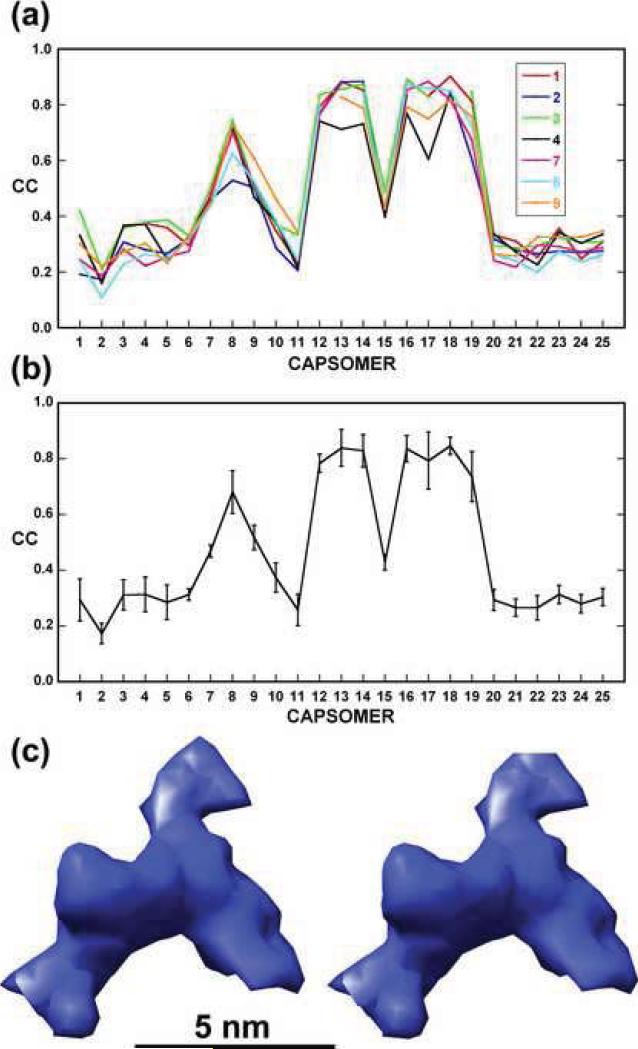
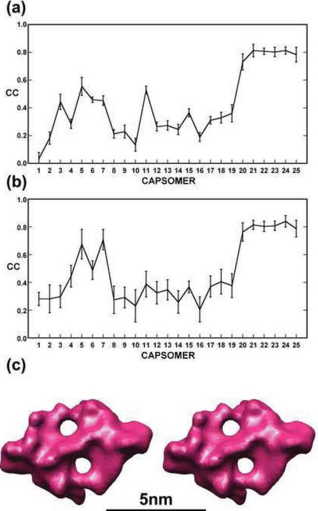
Chilo iridescent virus (CIV) is a large (approximately 1850 A diameter) insect virus with an icosahedral, T=147 capsid, a double-stranded DNA (dsDNA) genome, and an internal lipid membrane. The structure of CIV was determined to 13 A resolution by means of cryoelectron microscopy (cryoEM) and three-dimensional image reconstruction. A homology model of P50, the CIV major capsid protein (MCP), was built based on its amino acid sequence and the structure of the homologous Paramecium bursaria chlorella virus 1 Vp54 MCP. This model was fitted into the cryoEM density for each of the 25 trimeric CIV capsomers per icosahedral asymmetric unit. A difference map, in which the fitted CIV MCP capsomers were subtracted from the CIV cryoEM reconstruction, showed that there are at least three different types of minor capsid proteins associated with the capsomers outside the lipid membrane. "Finger" proteins are situated at many, but not all, of the spaces between three adjacent capsomers within each trisymmetron, and "zip" proteins are situated between sets of three adjacent capsomers at the boundary between neighboring trisymmetrons and pentasymmetrons. Based on the results of segmentation and density correlations, there are at least eight finger proteins and three dimeric and two monomeric zip proteins in one asymmetric unit of the CIV capsid. These minor proteins appear to stabilize the virus by acting as intercapsomer cross-links. One transmembrane "anchor" protein per icosahedral asymmetric unit, which extends from beneath one of the capsomers in the pentasymmetron to the internal leaflet of the lipid membrane, may provide additional stabilization for the capsid. These results are consistent with the observations for other large, icosahedral dsDNA viruses that also utilize minor capsid proteins for stabilization and for determining their assembly.


Figures

Fig. 1
Fig. 1. 3D reconstruction of CIV at 13 Å resolution
(a). Micrograph of vitrified CIV virions suspended over a hole in a carbon support film. The core contents (dsDNA genome and DNA binding proteins) do not uniformly fill the volume enclosed by the capsid. (b) Shaded-surface representation of CIV 3D density map viewed along an icosahedral 2-fold axis. (c) Central cross section, one pixel thick, viewed as in (b). A lipid bilayer, ~40 Å thick, follows the inner contour and icosahedral symmetry of the capsid shell. (d) Magnified view of the region outlined in (b). A black ellipse marks the location of an icosahedral 2-fold axis and the thin white line indicates the cleavage plane between two trisymmetrons. (e) Magnified view of the pentamer complex at the 5-fold vertex. This complex is easily distinguished from the trimeric capsomers in that it is larger, has a small axial hole and lacks a fiber. (f) Magnified view of boxed region in (c). (g) Same as (f) with the pentamer complex, three capsomers and their fibers individually outlined.
Fig. 2
Fig. 2. Organization of trimeric capsomers in CIV
(a) The CIV capsid has twenty trisymmetrons (one shown in light blue and another in dark blue) and twelve pentasymmetrons (two shown in magenta). One triangular face of the icosahedron is outlined in green. The asymmetric unit of the capsid includes 24 1/3 trimers (numbering scheme is arbitrary). Only one monomer of capsomer 10 (at an icosahedral 3-fold axis) is included in the icosahedral asymmetric unit. (b) Schematic diagram of inter-capsomer interactions within one asymmetric unit. Each capsomer is represented as a pseudo-hexamer (three double jelly-rolls, each depicted as a pair of red-green dots connected by a thick line). Capsomers form three classes (I, II, and III; Table 1) of interactions with neighboring capsomers as indicated by single, double, and triple dashed lines. The black ellipse, triangle, and pentagon symbols mark the icosahedral 2-, 3-, and 5-fold axes, respectively. Diagram does not depict curvature present in the arrangement of capsomers in the CIV structure.
Fig. 3
Fig. 3. Transmembrane anchor protein beneath CIV pentasymmetrons
(a) An equatorial-section, one pixel thick, through the CIV density map showing the pentamer vertex complex (arrow head) and a portion of the anchor protein (arrow). The bilayer membrane sharply curves in this region. (b) Similar to (a), but showing a section (parallel to but displaced ~21 Å from the equatorial section) that reveals additional details of the anchor protein. Two stick-like densities are apparent. The longer of the two (arrow) crosses both leaflets of the bilayer, whereas the other stops at the outer leaflet.
Fig. 4
Fig. 4. Minor proteins in CIV
(a). Upper panel: Inside view of “-(P50,core)” map with membrane density removed for clarity, in stereo and along an icosahedral 2-fold axis, shows a complex distribution of repeating density features. Some are arranged in a hexagonal array under the trisymmetrons whereas others are associated with the pentasymmetrons. Lower panel: Same as upper panel but colored to highlight similarly shaped features that were identified as putative, unique minor proteins. Twenty-seven finger proteins (colored blue, numbered 1 to 9 in the icosahedral asymmetric unit) form a tight network underneath each trisymmetron and their finger-like protrusions point towards the lipid membrane. Six zip dimers (red) and four zip monomers (orange) lie below the interface between adjacent trisymmetrons. Transmembrane anchor proteins (green) are the most readily identified features located underneath the pentasymmetrons. (b) Schematic, planar diagram of region depicted in (a), with the pentamer complex omitted for clarity. The ellipse, triangle, and pentagon symbols highlight the positions of 2-, 3-, and 5-fold icosahedral axes, respectively. All P50 trimers are depicted as three filled disks enclosed by a hexagon. The icosahedral asymmetric unit contains 24 1/3 of these capsomers (numbered in black). Each trisymmetron contains 55 capsomers, all oriented similarly and rotated by 60° relative to those in the adjacent trisymmetron. Twenty-seven finger proteins (blue) bind to each trisymmetron (nine within one asymmetric unit are numbered in white). Eighteen zip dimers (red) form the interface between one trisymmetron and its three adjacent trisymmetrons. Six zip monomers (orange) form a portion of the interface between a trisymmetron and its neighboring pentasymmetrons. The transmembrane anchor proteins (green) are associated with capsomers 2 and 3 beneath the pentasymmetron.
Fig. 5
Fig. 5. Correlation coefficient analysis and averaging of finger protein densities
(a) Plots of correlation coefficients computed between each of the seven, most prominent finger densities (1-4 and 7-9 in Fig. 4) and the corresponding densities at each of the other 24 capsomer sites. Densities associated with capsomers 12-14 and 16-19 show the highest correlations (average = 0.81). This analysis also indicates a high correlation for capsomer 8 (CC = 0.68) and capsomer 9 has the next highest correlation (CC = 0.52). (b) Same as (a) but only plotting the average correlation and its standard deviation (vertical bar) at each capsomer site. (c) Stereo view of the average density for the seven prominent finger proteins.
Fig. 6
Fig. 6. Correlation coefficient analysis and averaging of zip protein densities
(a) Plot of average correlation coefficient computed between each of the six, prominent densities representing zip dimers and the corresponding densities at each of the other 24 capsomer sites. Densities associated with capsomers 20-25 show the highest correlations (average = 0.79). (b) Same as (a) for density of a zip monomer. Two zip monomers (associated with capsomers 5 and 7) have a correlation greater than 0.67. (c) Stereo view of the average density for the six prominent zip dimers. Note that no 2-fold symmetry was enforced during this averaging procedure.

References

    1. Iyer LM, Aravind L, Koonin EV. Common origin of four diverse families of large eukaryotic DNA viruses. J. Virol. 2001;75:11720–11734. - PMC - PubMed
    1. Van Etten JL, Graves MV, MÜller DG, Boland W, Delaroque N. Phycodnaviridae - large DNA algal viruses. Arch. Virology. 2002;147:1479–1516. - PubMed
    1. La Scola B, Audic S, Robert C, Jungang L, de Lamballerie X, Drancourt M, Birtles R, Claverie JM, Raoult D. A giant virus in amoebae. Science. 2003;299:2033. - PubMed
    1. Zauberman N, Mutsafi Y, Halevy DB, Shimoni E, Klein E, Xiao C, Sun S, Minsky A. Distinct DNA exit and packaging portals in the virus Acanthamoeba polyphaga mimivirus. PLoS Biol. 2008;6:1104–1114. - PMC - PubMed
    1. Yan X, Chipman PR, Castberg T, Bratbak G, Baker TS. The marine algal virus PpV01 has an icosahedral capsid with T=219 quasisymmetry. J. Virol. 2005;79:9236–9243. - PMC - PubMed

Publication types