

Regional white matter volume differences in nondemented aging and Alzheimer's disease
- PMID: 19027860
- PMCID: PMC2810540
- DOI: 10.1016/j.neuroimage.2008.10.030
Regional white matter volume differences in nondemented aging and Alzheimer's disease
Abstract
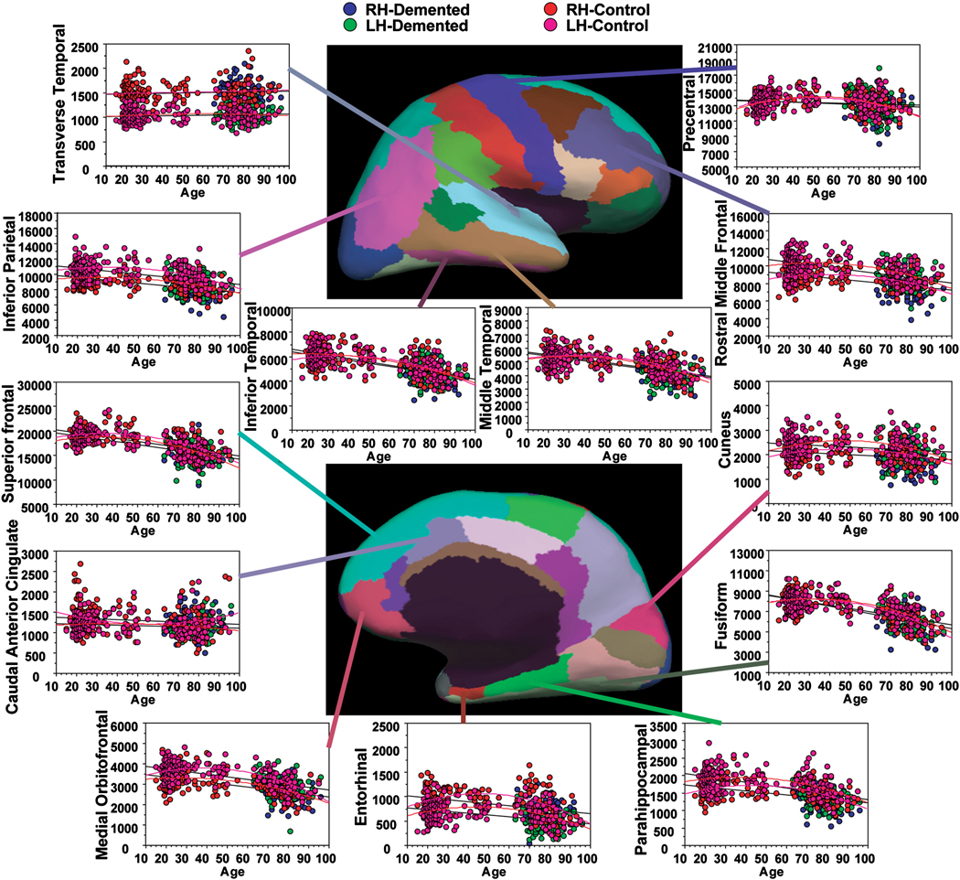
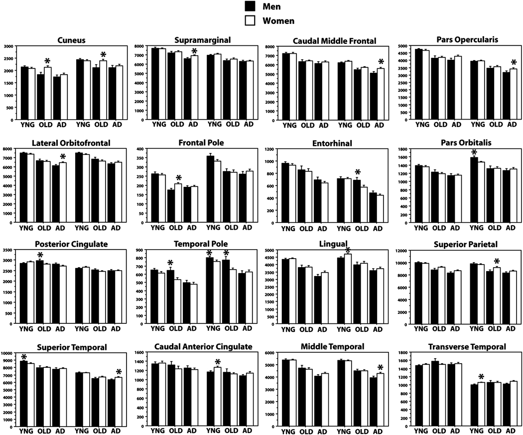
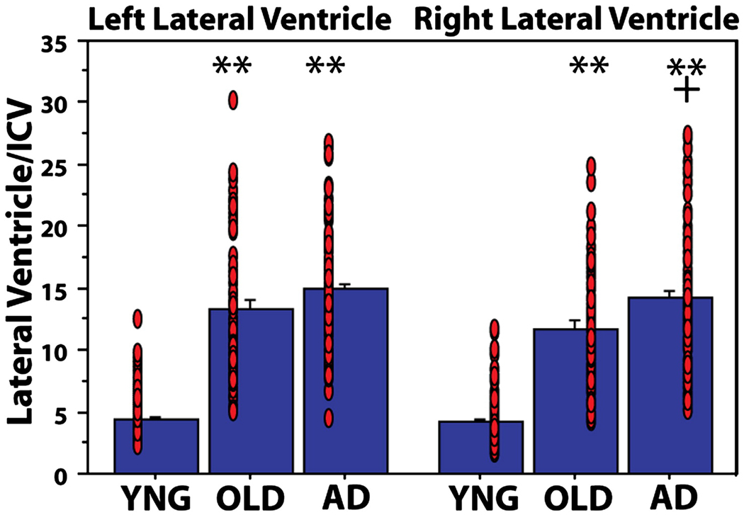
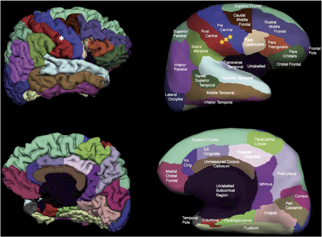
Accumulating evidence suggests that altered cerebral white matter (WM) influences normal aging, and further that WM degeneration may modulate the clinical expression of Alzheimer's disease (AD). Here we conducted a study of differences in WM volume across the adult age span and in AD employing a newly developed, automated method for regional parcellation of the subcortical WM that uses curvature landmarks and gray matter (GM)/WM surface boundary information. This procedure measures the volume of gyral WM, utilizing a distance constraint to limit the measurements from extending into the centrum semiovale. Regional estimates were first established to be reliable across two scan sessions in 20 young healthy individuals. Next, the method was applied to a large clinically-characterized sample of 299 individuals including 73 normal older adults and 91 age-matched participants with very mild to mild AD. The majority of measured regions showed a decline in volume with increasing age, with strong effects found in bilateral fusiform, lateral orbitofrontal, superior frontal, medial orbital frontal, inferior temporal, and middle temporal WM. The association between WM volume and age was quadratic in many regions suggesting that WM volume loss accelerates in advanced aging. A number of WM regions were further reduced in AD with parahippocampal, entorhinal, inferior parietal and rostral middle frontal WM showing the strongest AD-associated reductions. There were minimal sex effects after correction for intracranial volume, and there were associations between ventricular volume and regional WM volumes in the older adults and AD that were not apparent in the younger adults. Certain results, such as the loss of WM in the fusiform region with aging, were unexpected and provide novel insight into patterns of age associated neural and cognitive decline. Overall, these results demonstrate the utility of automated regional WM measures in revealing the distinct patterns of age and AD associated volume loss that may contribute to cognitive decline.
Figures

References
-
- Anderson JM, Hubbard BM, Coghill GR, Slidders W. The effect of advanced old age on the neurone content of the cerebral cortex. Observations with an automatic image analyser point counting method. J Neurol Sci. 1983;58:235–246. - PubMed
-
- Bartzokis G, Beckson M, Lu PH, Nuechterlein KH, Edwards N, Mintz J. Age-related changes in frontal and temporal lobe volumes in men: a magnetic resonance imaging study. Arch Gen Psychiatry. 2001;58:461–465. - PubMed
-
- Bartzokis G, Cummings JL, Sultzer D, Henderson VW, Nuechterlein KH, Mintz J. White matter structural integrity in healthy aging adults and patients with Alzheimer disease: a magnetic resonance imaging study. Arch Neurol. 2003;60:393–398. - PubMed
Publication types
MeSH terms
Grants and funding
- NR010827/NR/NINR NIH HHS/United States
- R01 NS039581/NS/NINDS NIH HHS/United States
- AG05886/AG/NIA NIH HHS/United States
- P50 AG005681/AG/NIA NIH HHS/United States
- P41 RR014075/RR/NCRR NIH HHS/United States
- K01 AG024898/AG/NIA NIH HHS/United States
- AG05681/AG/NIA NIH HHS/United States
- HHMI/Howard Hughes Medical Institute/United States
- NS39581/NS/NINDS NIH HHS/United States
- RR14075/RR/NCRR NIH HHS/United States
- P41RR14075/RR/NCRR NIH HHS/United States
- R01 AG034556/AG/NIA NIH HHS/United States
- F32 AG005886/AG/NIA NIH HHS/United States
- R01 NR010827/NR/NINR NIH HHS/United States
- AG024898/AG/NIA NIH HHS/United States
LinkOut - more resources
Full Text Sources
Medical
