. 2009 Jan 16;378(3):433-8.
doi: 10.1016/j.bbrc.2008.11.050. Epub 2008 Nov 24.

Proteomic profiling of human plasma exosomes identifies PPARgamma as an exosome-associated protein

Affiliations

Proteomic profiling of human plasma exosomes identifies PPARgamma as an exosome-associated protein

Christopher Looze et al. Biochem Biophys Res Commun. .

Abstract

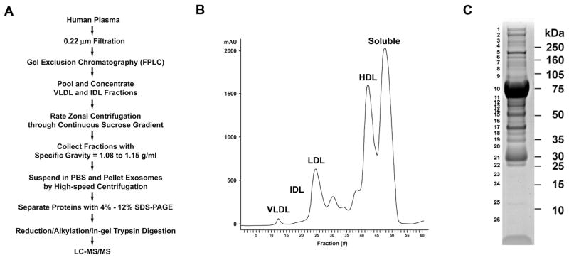
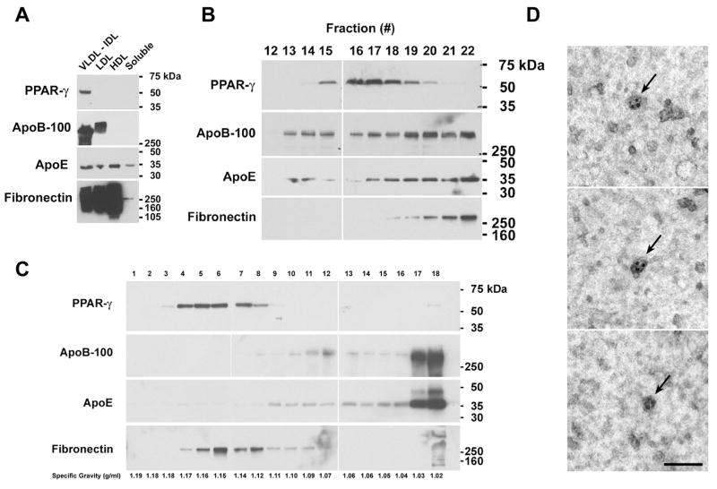
Exosomes are nanovesicles that are released from cells as a mechanism of cell-free intercellular communication. Only a limited number of proteins have been identified from the plasma exosome proteome. Here, we developed a multi-step fractionation scheme incorporating gel exclusion chromatography, rate zonal centrifugation through continuous sucrose gradients, and high-speed centrifugation to purify exosomes from human plasma. Exosome-associated proteins were separated by SDS-PAGE and 66 proteins were identified by LC-MS/MS, which included both cellular and extracellular proteins. Furthermore, we identified and characterized peroxisome proliferator-activated receptor-gamma (PPARgamma), a nuclear receptor that regulates adipocyte differentiation and proliferation, as well as immune and inflammatory cell functions, as a novel component of plasma-derived exosomes. Given the important role of exosomes as intercellular messengers, the discovery of PPARgamma as a component of human plasma exosomes identifies a potential new pathway for the paracrine transfer of nuclear receptors.


Figures

Figure 1
Figure 1. Isolation of Exosomes Co-segregating with the IDL and VLDL Fractions of Human Plasma for Proteomic Analysis
A. Flowchart for the isolation of exosomes that co-segregate with the IDL and VLDL fractions of human plasma. B. Chromatogram of fractions of human plasma separated by gel exclusion chromatography using FPLC. C. Image of Coomassie blue-stained 4%-12% 1D-SDS PAGE gel of exosomes isolated from human plasma. Molecular mass markers are shown on the right and gel slice numbers are shown on the left.
Figure 2
Figure 2. Characterization of PPARγ Exosomes that Co-segregate with the IDL Fraction of Human Plasma
A. Samples (20 μg) of FPLC fractions were immunoblotted and reacted with antibodies against PPARγ. Membranes were stripped and re-probed with antibodies against apolipoprotein B-100, apolipoprotein E, and fibronectin. This blot is representative of two independent experiments that demonstrated the same result. B. Proteins from individual tubes that corresponded to the VLDL and IDL fractions of human plasma were precipitated with 10% TCA, immunoblotted, and reacted with antibodies against PPARγ Membranes were stripped and re-probed with antibodies against apolipoprotein B-100, apolipoprotein E, and fibronectin. Individual tube numbers are shown on top. This blot is representative of five independent experiments that demonstrated the same result. C. A sample containing 435 μg of IDL proteins from human plasma was subjected to rate zonal centrifugation through a continuous sucrose gradient followed by immunoblotting. Lane numbers correspond to the fractions collected. Specific gravity of individual fractions are shown below. This blot is representative of two independent experiments that demonstrated the same result. D. Characterization by immunoelectron microscopy of PPARγ exosomes that co-segregate with the IDL fraction of human plasma. Exosomes from the IDL fraction of human plasma were concentrated and visualized by immunogold electron microscopy using antibodies that react with PPARγ. Three separate fields are shown. The arrows indicate PPARγ exosomes each labeled with 2–4 silver-enhanced gold particles (5–12 nm). The bar denotes 100 nm.

References

    1. Thery C, Zitvogel L, Amigorena S. Exosomes: composition, biogenesis and function. Nat Rev Immunol. 2002;2:569–579. - PubMed
    1. van Niel G, Porto-Carreiro I, Simoes S, Raposo G. Exosomes: a common pathway for a specialized function. J Biochem (Tokyo) 2006;140:13–21. - PubMed
    1. Caby MP, Lankar D, Vincendeau-Scherrer C, Raposo G, Bonnerot C. Exosomal-like vesicles are present in human blood plasma. Int Immunol. 2005;17:879–887. - PubMed
    1. Zhang J, Hawari FI, Shamburek RD, Adamik B, Kaler M, Islam A, Liao DW, Rouhani FN, Ingham M, Levine SJ. Circulating TNFR1 exosome-like vesicles partition with the LDL fraction of human plasma. Biochem Biophys Res Commun. 2008;366:579–584. - PMC - PubMed
    1. Wang G, Wu WW, Zeng W, Chou CL, Shen RF. Label-free protein quantification using LC-coupled ion trap or FT mass spectrometry: Reproducibility, linearity, and application with complex proteomes. J Proteome Res. 2006;5:1214–1223. - PubMed

Publication types

LinkOut - more resources