Review
. 2009 Mar;33(3):367-82.
doi: 10.1016/j.neubiorev.2008.10.009. Epub 2008 Nov 6.

A developmental neurobiological model of motivated behavior: anatomy, connectivity and ontogeny of the triadic nodes

Affiliations
Review

A developmental neurobiological model of motivated behavior: anatomy, connectivity and ontogeny of the triadic nodes

Monique Ernst et al. Neurosci Biobehav Rev. 2009 Mar.

Abstract

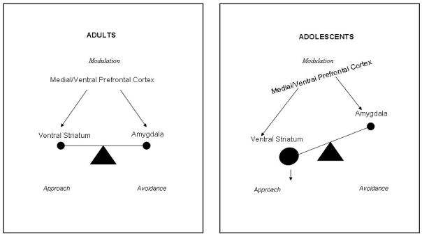
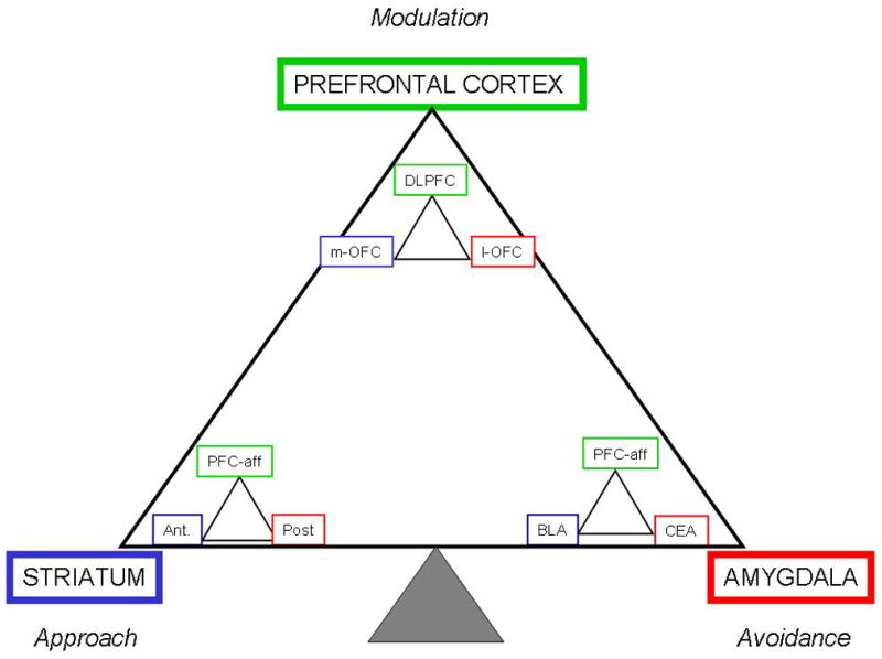
Adolescence is the transition period that prepares individuals for fulfilling their role as adults. Most conspicuous in this transition period is the peak level of risk-taking behaviors that characterize adolescent motivated behavior. Significant neural remodeling contributes to this change. This review focuses on the functional neuroanatomy underlying motivated behavior, and how ontogenic changes can explain the typical behavioral patterns in adolescence. To help model these changes and provide testable hypotheses, a neural systems-based theory is presented. In short, the Triadic Model proposes that motivated behavior is governed by a carefully orchestrated articulation among three systems, approach, avoidance and regulatory. These three systems map to distinct, but overlapping, neural circuits, whose representatives are the striatum, the amygdala and the medial prefrontal cortex. Each of these system-representatives will be described from a functional anatomy perspective that includes a review of their connectivity and what is known of their ontogenic changes.


Figures

Figure 1
Figure 1
Triadic Model. This model is a heuristic representation of a neural systems approach to the neurobiology of motivated behavior. Generically, behavior results from the integration of signals underlying the coding of approach, the coding of avoidance and the reciprocal modulation of both latter signals. Taken as the gold standard, the adult pattern is shown as a balanced system. Compared to this balanced system, the pattern expected in adolescents is tilted towards approach behavior. In this model, the representatives of the three systems include amygdala for avoidance, striatum for approach and medial prefrontal cortex for modulation. Arrows represent relative medial prefrontal cortical control over striatum and amygdala rather than the array of anatomic connections.
Figure 2
Figure 2
AD. Rendering of the approximate boundaries for the amygdala (orange), hippocampus (red) and striatopallidal structures (yellow) on a 3-D structural MRI. The boundaries of amygdala and hippocampal subregions cannot be reliably resolved; therefore the images are drawn based on the location of these structures in human postmortem sections. The planes intersect at the following Talairach coordinates x=20mm, y=−16mm and z=−2mm. On the sagittal view (A.), the location of the extended amygdala is represented as an orange strip bridging the dorsal amygdala and bed nucleus of the stria terminalis. Both the medial or central extended amygdala follow this rostrocaudal trajectory. The axial view (B.) is from a plane that passes right below the striatum and the bed nucleus of the amygdala, and reveals the amygdala (orange) positioned anteriorly to the hippocampus (red). Both structures form the medial wall of the temporal horn of the lateral ventricle. The coronal view (C.) delineates the putamen (bright yellow). The medial (red with black outline) and central extended amygdala (orange with black outline) are illustrated on the left hemisphere. The superior part of the bed nucleus of the stria terminalis is also being represented at the inferior part of the caudate nucleus (orange circle). In D., this area (boxed D.) is enlarged to provide a better vision of the relative location of these structures. MEA = Medial Extended Amygdala; CEA = Central Extended Amygdala.
Figure 3
Figure 3
Simplified rendering of the general locations of amygdala subregions superimposed on a coronal section at the level of the amygdalohippocampal transition (AHA). The main basolateral nuclear group (BLNG, in tan) sends excitatory inputs to the CeN. These excitatory inputs also collateralize to innervate the GABAergic intercalated neurons (blue dots), which inhibit CeN activity. This arrangement provides a means for feedforward inhibitory modulation of CeN outputs. The intercalated islands are also importantly innervated by PFC inputs (see text). AStr= amygdalostriatal area, BLNG= basolateral nuclear group, CeN= central nucleus, MeN= medial nucleus
Figure 4
Figure 4
Representation of the projections of the amygdala (blue) and cortico-striato-pallido-thalamo-cortical loops (black). The hatched lines signify that these connections are not in this sagittal plane, but go more medially. The solid lines are in the plane of the section. The amygdala projection to the ventral striatum is the only one that is unidirectional (green arrow). All other amygdala projections are bidirectional (blue) OMPFC= orbito-medial prefrontal cortex; ant. Insula = anterior insula; BA 25 = Brodmann area 25; Pal. = pallidum; PFC = prefrontal cortex; Thal. = thalamus; Str. = striatum
Figure 5
Figure 5
Fractal Triadic Model. The fractal Triadic Model is a more comprehensive representation of the Triadic Model, because it takes into account the heteromodal role of each of the nodes of the triad. Each of the nodes of approach, avoidance and regulatory control is itself the seat of a triadic formation that mirrors the overall organization. Examples of regions previously identified to be associated in some studies with either approach or avoidance responses are indicated. However, similarly to the original Triadic Model, this representation is highly schematic and not all-inclusive. DLPFC=dorsolateral prefrontal cortex, m-OFC=medial orbital frontal cortex, l-OFC lateral orbital frontal cortex, PFC-aff, prefrontal cortical afferents, Ant=anterior striatum, Post=posterior striatum, BLA=basolateral amygdala, CEA=central amygdala

References

    1. Adam J, Baulac M, Hauw JJ, Laplane D, Duyckaerts C. Behavioral symptoms after pallido-nigral lesions: a clinico-pathological case. Neurocase. 2008;14:125–130. - PubMed
    1. Adolphs R, Gosselin F, Buchanan TW, Tranel D, Schyns P, Damasio AR. A mechanism for impaired fear recognition after amygdala damage. Nature. 2005;433:68–72. - PubMed
    1. Adolphs R, Tranel D, Damasio H, Damasio A. Impaired recognition of emotion in facial expressions following bilateral damage to the human amygdala. Nature. 1994;372:669–672. - PubMed
    1. Adolphs R, Tranel D, Damasio H, Damasio AR. Fear and the human amygdala. J Neurosci. 1995;15:5879–5891. - PMC - PubMed
    1. Aggleton JP. The functional effects of amygdala lesions in humans: A comparison with findings from monkeys. In: Aggleton JP, editor. The amygdala: Neurobiological aspects of emotion, memory, and mental dysfunction. Wiley-Liss Inc.; New York: 1992. pp. 485–503.