. 2009 Jan 23;284(4):2576-83.
doi: 10.1074/jbc.M804394200. Epub 2008 Nov 20.

Functional UDP-xylose transport across the endoplasmic reticulum/Golgi membrane in a Chinese hamster ovary cell mutant defective in UDP-xylose Synthase

Affiliations

Functional UDP-xylose transport across the endoplasmic reticulum/Golgi membrane in a Chinese hamster ovary cell mutant defective in UDP-xylose Synthase

Hans Bakker et al. J Biol Chem. .

Abstract

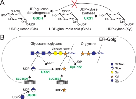
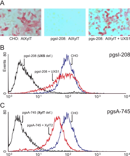
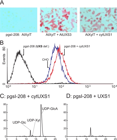
In mammals, xylose is found as the first sugar residue of the tetrasaccharide GlcAbeta1-3Galbeta1-3Galbeta1-4Xylbeta1-O-Ser, initiating the formation of the glycosaminoglycans heparin/heparan sulfate and chondroitin/dermatan sulfate. It is also found in the trisaccharide Xylalpha1-3Xylalpha1-3Glcbeta1-O-Ser on epidermal growth factor repeats of proteins, such as Notch. UDP-xylose synthase (UXS), which catalyzes the formation of the UDP-xylose substrate for the different xylosyltransferases through decarboxylation of UDP-glucuronic acid, resides in the endoplasmic reticulum and/or Golgi lumen. Since xylosylation takes place in these organelles, no obvious requirement exists for membrane transport of UDP-xylose. However, UDP-xylose transport across isolated Golgi membranes has been documented, and we recently succeeded with the cloning of a human UDP-xylose transporter (SLC25B4). Here we provide new evidence for a functional role of UDP-xylose transport by characterization of a new Chinese hamster ovary cell mutant, designated pgsI-208, that lacks UXS activity. The mutant fails to initiate glycosaminoglycan synthesis and is not capable of xylosylating Notch. Complementation was achieved by expression of a cytoplasmic variant of UXS, which proves the existence of a functional Golgi UDP-xylose transporter. A approximately 200 fold increase of UDP-glucuronic acid occurred in pgsI-208 cells, demonstrating a lack of UDP-xylose-mediated control of the cytoplasmically localized UDP-glucose dehydrogenase in the mutant. The data presented in this study suggest the bidirectional transport of UDP-xylose across endoplasmic reticulum/Golgi membranes and its role in controlling homeostasis of UDP-glucuronic acid and UDP-xylose production.


Figures

FIGURE 1.
FIGURE 1.
UDP-xylose metabolism in mammalian cells. A, UDP-Xyl is synthesized in two steps from UDP-Glc by the enzymes UGDH, forming UDP-GlcA, and UXS, also referred to as UDP-glucuronic acid decarboxylase. UGDH is inhibited by the product of the second enzyme, UDP-Xyl (42). B, in mammals, UDP-Xyl is synthesized within the lumen of the ER/Golgi, where it is substrate for different xylosyltransferases incorporating xylose in the glycosaminoglycan core (XylT1 and XylT2) or in O-glucose-linked glycans. The nucleotide sugar transporter SLC35D1 (52) has been shown to transport UDP-GlcA over the ER membrane and SLC35B4 (29) to transport UDP-Xyl over the Golgi membrane. The function of this latter transporter is unclear.
FIGURE 2.
FIGURE 2.
Complementation of pgsI-208 by UXS. A, CHO or pgsI-208 cells transfected with AtXylT. The product of AtXylT, xylosylated N-glycans, was detected on the cell surface using anti-HRP, reactive to xylosylated N-glycans of this protein (40). B and C, pgsI-208 and pgsA-745 cells were stained with antibody RB4Ea12 (35, 36) against heparan sulfate and analyzed by flow cytometry. B, CHO cells (blue) compared with pgsI-208 (black) and pgsI-208 stably transfected with human UXS1 (red). C, CHO cells (blue) compared with xylosyltransferase-deficient mutant pgsA-745 (black) and pgsA-745 stably transfected with human XylT2 (red).
FIGURE 3.
FIGURE 3.
Glycan analysis of Notch EGF1-5 by mass spectrometry. pgsA-745 and pgsI-208 cells were transiently transfected with a construct encoding EGF repeats 1-5 of mouse Notch1. Tryptic peptides of the purified secreted product were analyzed by liquid chromatography-MS/MS for the presence of peptides modified with O-glucose. A, identification of the O-glucose trisaccharide form of a peptide from the pgsA-745 sample. The top panel shows an MS spectrum of material eluting at 52.9 min. The ions labeled [M + 3H]3+ and [M + 4H]4+ match the predicted mass for triply and quadruply charged forms of the O-glucose trisaccharide form of 137SCQQADPCASNPCANGGQCLPFESSYICR165, a tryptic peptide from EGF 4 previously demonstrated to be modified with O-glucose trisaccharide.3 Collision-induced dissociation fragmentation of the triply charged form of this peptide resulted in the MS/MS spectrum shown in the bottom panel. The major product ions at m/z 1210.9, 1167.1, and 1112.9 correspond to sequential losses of a pentose, a second pentose, and a hexose. Numerous sequence fragment ions (y-ions are shown) are observed that confirm the identity of the peptide. Ions selected for fragmentation in the MS spectrum are identified by red diamonds. The position of the parent ion fragmented in the MS/MS spectrum is identified with a blue diamond. B, identification of the O-glucose monosaccharide form of the peptide from the pgsI-208 sample. The top panel shows an MS spectrum of material eluting at 53.7 min, slightly later than the trisaccharide form (consistent with the loss of two hydrophilic xylose residues). The ions labeled [M + 3H]3+ and [M + 4H]4+ match the predicted mass for triply and quadruply charged forms of the glycopeptide. Collision-induced dissociation fragmentation of the triply charged form resulted in the MS/MS spectrum shown in the bottom panel. The major product ion, m/z 1112.8, matches the mass for the unglycosylated peptide. C, the trisaccharide form is present in samples from pgsA-745 but not pgsI-208. The data from both samples was searched for the ion corresponding to the triply charged form of the trisaccharide form, m/z 1255.1 (see A). D, the monosaccharide form is present in samples from psgI-208 but not pgsA-745. The data from both samples was searched for the ion corresponding to the triply charged form of the monosaccharide form, m/z 1166.7 (see B).
FIGURE 4.
FIGURE 4.
UDP-Glc, UDP-GlcA, and UDP-Xyl content of CHO cells. Nucleotide sugars of wild-type, pgsA-745, and pgsI-208 cells were separated in a two-step HPLC as described under “Experimental Procedures” (A-C). Fractions of the first HPLC run were collected, and combined fractions of UDP-Glc, UDP-GlcA, and UDP-Xyl (based on the position of standards (D)) were applied to a second run. E shows the pgsI-208 profile on a decreased scale to demonstrate the dramatic increase of UDP-GlcA compared with UDP-Glc. UDP-GlcA in pgsI-208 is estimated to be increased by a factor of 200 over wild type CHO cell levels based on peak area. *, location of UDP-Xyl as confirmed by mass spectrometry as described (not shown) (39).
FIGURE 5.
FIGURE 5.
Complementation of pgsI-208 by cytoplasmic expression of UXS. A, pgsI-208 cells were transfected with AtXylT or cotransfected with AtUXS3 or cytUXS1 and stained with anti-HRP as in Fig. 2A. B, cells were stained with the anti-heparan sulfate antibody as in Fig. 2B and analyzed by flow cytometry. CHO cells (blue) compared with pgsI-208 (black) and pgsI-208 stably transfected with human UXS1 expressed in the cytoplasm (red). C and D, nucleotide sugar composition analysis (as in Fig. 4) of pgsI-208 expressing human cytUXS1 (C) or UXS1 (D). Identity of the UDP-Xyl peak has been confirmed by mass spectrometry.

References

    1. Roden, L., and Armand, G. (1966) J. Biol. Chem. 241 65-70 - PubMed
    1. Hase, S., Kawabata, S., Nishimura, H., Takeya, H., Sueyoshi, T., Miyata, T., Iwanaga, S., Takao, T., Shimonishi, Y., and Ikenaka, T. (1988) J. Biochem. (Tokyo) 104 867-868 - PubMed
    1. Moloney, D. J., Shair, L. H., Lu, F. M., Xia, J., Locke, R., Matta, K. L., and Haltiwanger, R. S. (2000) J. Biol. Chem. 275 9604-9611 - PubMed
    1. Ponighaus, C., Ambrosius, M., Casanova, J. C., Prante, C., Kuhn, J., Esko, J. D., Kleesiek, K., and Götting, C. (2007) J. Biol. Chem. 282 5201-5206 - PubMed
    1. Voglmeir, J., Voglauer, R., and Wilson, I. B. (2007) J. Biol. Chem. 282 5984-5990 - PMC - PubMed

Publication types

LinkOut - more resources