

Altered plasticity of the parasympathetic innervation in the recovering rat submandibular gland following extensive atrophy
- PMID: 19028809
- PMCID: PMC2773434
- DOI: 10.1113/expphysiol.2008.045112
Altered plasticity of the parasympathetic innervation in the recovering rat submandibular gland following extensive atrophy
Abstract
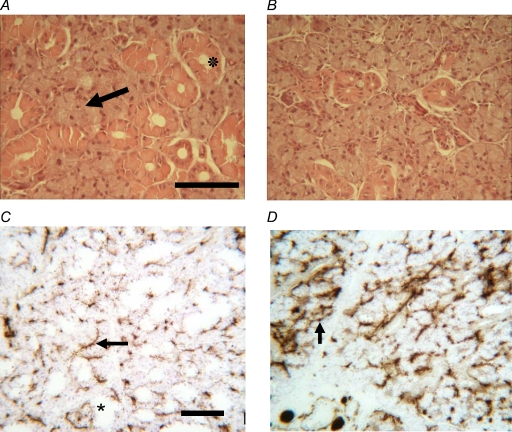
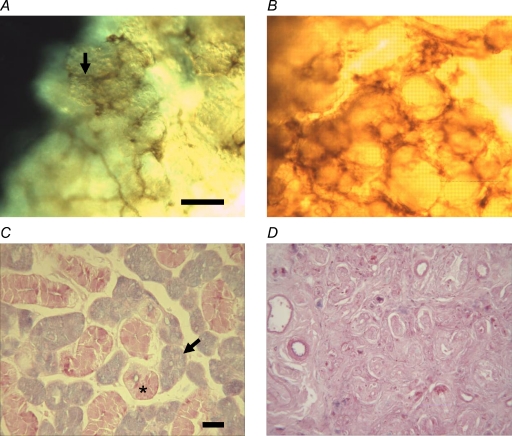
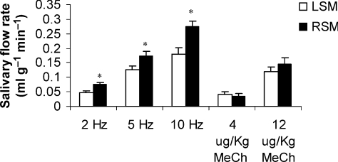
Adult rat submandibular glands have a rich autonomic innervation, with parasympathetic and sympathetic nerves working in synergy rather than antagonistically. Ligation of the secretory duct rapidly causes atrophy and the loss of most acini, which are the main target cell for parasympathetic nerves. Following deligation, there is a recovery of gland structure and function, as assessed by autonomimetic stimulation. This study examines whether the parasympathetic nerves reattach to new target cells to form functional neuro-effector junctions. Under recovery anaesthesia, the submandibular duct of adult male rats was ligated via an intra-oral approach to avoid damaging the chorda-lingual nerve. Four weeks later, rats were either killed or anaesthetized and the ligation clip removed. Following a further 8 weeks, both submandibular ducts were cannulated under terminal anaesthesia. Salivary flows were then stimulated electrically (chorda-lingual nerve at 2, 5 and 10 Hz) and subsequently by methacholine (whole-body infusion at two doses). Glands were excised, weighed and divided for further in vitro studies or fixed for histological examination. Ligation of ducts caused 75% loss of gland weight, with the loss of most acinar cells. Of the remaining acini, only 50% were innervated despite unchanged choline acetyltransferase activity, suggesting few parasympathetic nerves had died. Following deligation, submandibular glands recovered half their weight and had normal morphology. Salivary flows from both glands (per unit of gland tissue) were similar when evoked by methacholine but greater from the deligated glands when evoked by nerve stimulation. This suggests that parasympathetic nerves had reattached to new target cells in the recovered glands at a greater ratio than normal, confirming reinnervation of the regenerating gland.
Figures
References
-
- Banns HE, Ekström J, Mann SP. Effects of duct ligation on choline acetyltransferase activity in salivary glands of rats. Acta Physiol Scand. 1979;106:431–435. - PubMed
-
- Bogart BI. Fine structural localization of cholinesterase activity in the rat submandibular gland. J Histochem Cytochem. 1970;18:730–739. - PubMed
-
- Carpenter GH, Proctor GB, Ebersole LE, Garrett JR. Secretion of IgA by rat parotid and submandibular cells in response to autonomimetic stimulation in vitro. Int Immunopharmacol. 2004;4:1005–1014. - PubMed
Publication types
MeSH terms
Substances
Grants and funding
LinkOut - more resources
Full Text Sources