. 2008 Dec 2;105(48):18913-8.
doi: 10.1073/pnas.0810097105. Epub 2008 Nov 21.

Spontaneous induction of murine pancreatic intraepithelial neoplasia (mPanIN) by acinar cell targeting of oncogenic Kras in adult mice

Affiliations

Spontaneous induction of murine pancreatic intraepithelial neoplasia (mPanIN) by acinar cell targeting of oncogenic Kras in adult mice

Nils Habbe et al. Proc Natl Acad Sci U S A. .

Abstract

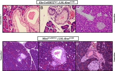
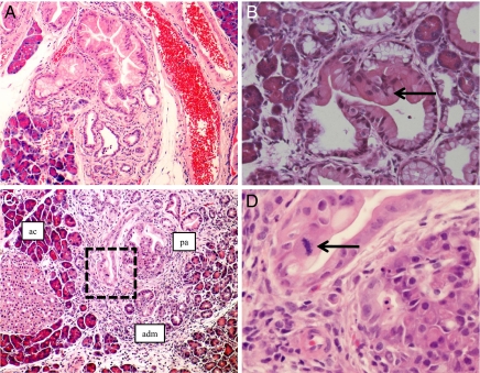
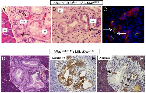
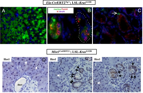
Pancreatic ductal adenocarcinoma (PDAC) is believed to arise through a multistep model comprised of putative precursor lesions known as pancreatic intraepithelial neoplasia (PanIN). Recent genetically engineered mouse models of PDAC demonstrate a comparable morphologic spectrum of murine PanIN (mPanIN) lesions. The histogenesis of PanIN and PDAC in both mice and men remains controversial. The most faithful genetic models activate an oncogenic Kras(G12D) knockin allele within the pdx1- or ptf1a/p48-expression domain of the entire pancreatic anlage during development, thus obscuring the putative cell(s)-of-origin from which subsequent mPanIN lesions arise. In our study, activation of this knockin Kras(G12D) allele in the Elastase- and Mist1-expressing mature acinar compartment of adult mice resulted in the spontaneous induction of mPanIN lesions of all histological grades, although invasive carcinomas per se were not seen. We observed no requirement for concomitant chronic exocrine injury in the induction of mPanIN lesions from the mature acinar cell compartment. The acinar cell derivation of the mPanINs was established through lineage tracing in reporter mice, and by microdissection of lesional tissue demonstrating Cre-mediated recombination events. In contrast to the uniformly penetrant mPanIN phenotype observed following developmental activation of Kras(G12D) in the Pdx1-expressing progenitor cells, the Pdx1-expressing population in the mature pancreas (predominantly islet beta cells) appears to be relatively resistant to the effects of oncogenic Kras. We conclude that in the appropriate genetic context, the differentiated acinar cell compartment in adult mice retains its susceptibility for spontaneous transformation into mPanIN lesions, a finding with potential relevance vis-à-vis the origins of PDAC.


Conflict of interest statement

The authors declare no conflict of interest.

Figures

Fig. 1.
Fig. 1.
Targeting KrasG12D to mature acinar cells results in spontaneous development of murine PanIN (mPanIN) lesions. (A) Low-grade mPanIN lesion (mPanIN-1A) in an Ela-CreERT2Tg/+; LSL-KrasG12D mouse, at 2 months posttamoxifen induction. (B) A second example of a low-grade mPanIN lesion (mPanIN-1A) in an Ela-CreERT2Tg/+; LSL-KrasG12D mouse. Features of low-grade mPanIN lesions illustrated in A and B includex the basally located nuclei, retained nuclear polarity, and absence of nuclear pleomorphism or mitoses. In addition, the abundant intracellular mucin is a characteristic feature. Note the absence of either lobular atrophy or histological evidence of pancreatitis in the surrounding parenchyma. (C) Absence of a discernible pancreatic phenotype in a noninduced Ela-CreERT2Tg/+; LSL-KrasG12D mouse, at 12 months of age. (D) Low-grade mPanIN lesion (mPanIN-1A) in a Mist1CreERT2/+; LSL-KrasG12D mouse, at 2 months posttamoxifen induction. The overall histological features are almost identical to those observed in the Ela-CreERT2Tg/+; LSL-KrasG12D mice, including the absence of either lobular atrophy or histological evidence of pancreatitis in the surrounding parenchyma. (E) A second example of a low-grade mPanIN lesion (mPanIN-1A) in a Mist1CreERT2/+; LSL-KrasG12D mouse, at 2 months post-tamoxifen induction. The diagnostic features of low-grade mPanIN are present. (F) Absence of a discernible pancreatic phenotype in a noninduced Mist1CreERT2/+; LSL- KrasG12D mouse, at 12 months of age.
Fig. 2.
Fig. 2.
The entire histological spectrum of mPanINs is observed with acinar targeting of mutant KrasG12D. (A) An example of high-grade mPanIN lesion (mPanIN-2) in the pancreas of an Ela-CreERT2Tg/+; LSL-KrasG12D mouse, harvested at 12 months posttamoxifen induction. The mPanIN-2 arises on the backdrop of lobulocentric trophy, while normal acinar parenchyma is seen toward the periphery. (B) An example of high-grade mPanIN lesion (mPanIN-2) in the pancreas of a Mist1CreERT2/+; LSL-KrasG12D mouse, harvested at 12 months post-tamoxifen induction. Micropapillary architecture with loss nuclear polarity is discernible (arrow). (C) An example of the highest grade mPanIN lesion (mPanIN-3, or carcinoma-in situ) in an Ela-CreERT2Tg/+; LSL-KrasG12D mouse, at 12 months posttamoxifen induction. Areas of histologically normal acinar parenchyma (ac) merge into acinar ductal metaplasia (adm), which in turn, transition into the high-grade mPanIN lesion (pa). (D) High-power view of the boxed area within the mPanIN-3 lesion illustrated in B, demonstrating the presence of a mitotic figure (arrow), nuclear pleomorphism, and loss of nuclear polarity.
Fig. 3.
Fig. 3.
Acinar-ductal metaplasia and biphenotypic differentiation are observed in acinar cell-derived mPanIN lesions. (A) Histological sections of pancreata from an Ela-CreERT2Tg/+; LSL-KrasG12D mouse at 6 months post-tamoxifen induction demonstrate acinar-ductal metaplastic lesions (adm) bridging the parenchyma between normal acinar structures (ac) and fully developed mPanINs (pa). The metaplastic structures contain differentiated acinar cells, as evidenced by their coarse zymogen granules (arrows), intermixed with mucinous epithelium characteristic of low-grade mPanIN lesions. (B) Another representative example of acinar ductal metaplasia (adm) in an Ela-CreERT2Tg/+; LSL-KrasG12D mouse at 10 months post-tamoxifen induction, transitioning into a more obvious mPanIN lesion (pa) towards the left of the field. Single acinar cells (arrows) are seen within the metaplastic epithelium. (C) Immunofluorescence with anti-amylase antibody (green channel) confirms the presence of residual acinar cells within metaplastic epithelium (anti-Ecad = red channel). DAPI (blue channel) is used as nuclear counterstain. Pancreatic section from an Ela-CreERT2Tg/+; LSL-KrasG12D mouse at 6 months post-tamoxifen induction. (D) A fully developed mPanIN-2 lesion in a Mist1CreERT2/+; LSL-KrasG12D mouse, at 4 months of age. (E) Immunophenotypic analysis of the mPanIN lesion illustrated in D shows extensive labeling with cytokeratin CK19, a marker of ductal differentiation. (F) Immunophenotypic analysis of the mPanIN lesion illustrated in D shows scattered labeling with amylase (arrows), consistent with biphenotypic acinar-ductal differentiation.
Fig. 4.
Fig. 4.
Activation of the Notch signaling pathway is an early molecular alteration in acinar-derived murine PanIN lesions. (A) No discernible expression of the Notch1 intracytoplasmic domain (N1-ICD, red channel) is seen in the uninvolved acinar parenchyma distant from mPanIN lesions, in a 4-month-old Ela-CreERT2Tg/+; LSL-KrasG12D mouse. DAPI is used as nuclear counterstain (blue channel). (B) Strong nuclear and cytoplasmic expression of N1-ICD is seen in acinar ductal metaplasia from a 4-month-old Ela-CreERT2Tg/+; LSL-KrasG12D mouse. Intense nuclear N1-ICD is particularly prominent in the individual remnant acinar cells, as discernible by their persistent amylase expression, within the metaplastic structures (dotted line). (C) Diffuse cytoplasmic N1-ICD expression in fully formed mPanIN lesions in Ela-CreERT2Tg/+; LSL-KrasG12D mice. Single amylase-expressing acinar cells are evident within the mPanIN lesions (see also Fig. 3). (D) No nuclear Hes1 expression is seen in the uninvolved acinar parenchyma of a 2month old Mist1CreERT2/+; LSL-KrasG12D mouse and the only cells expressing the protein are centroacinar cells (white arrows). The ductal epithelium in the photomicrograph is also Hes1-negative. (E) Uniformly strong nuclear Hes1 expression is observed in acinar ductal metaplasia and incipient mPanIN lesions in a Mist1CreERT2/+; LSL-KrasG12D mouse (black arrows). In contrast, the adjacent uninvolved acinar parenchyma is negative sans the expected labeling in centroacinar cells (white arrows). (F) Additional examples of nuclear Hes1 labeling in acinar ductal metaplasia and incipient mPanIN lesions Mist1CreERT2/+; LSL-KrasG12D mice (black arrows), reiterating the absence of expression in adjacent uninvolved acinar cells.

References

    1. Hruban RH, et al. Pancreatic intraepithelial neoplasia: A new nomenclature and classification system for pancreatic duct lesions. Am J Surg Pathol. 2001;25:579–586. - PubMed
    1. Longnecker DS, Memoli V, Pettengill OS. Recent results in animal models of pancreatic carcinoma: Histogenesis of tumors. Yale J Biol Med. 1992;65:457–465. - PMC - PubMed
    1. Pour PM, Pandey KK, Batra SK. What is the origin of pancreatic adenocarcinoma? Mol Cancer. 2003;2:13. - PMC - PubMed
    1. Jamieson JD. Prospectives for cell and organ culture systems in the study of pancreatic carcinoma. J Surg Oncol. 1975;7:139–141. - PubMed
    1. Cubilla AL, Fitzgerald PJ. Morphological lesions associated with human primary invasive nonendocrine pancreas cancer. Cancer Res. 1976;36:2690–2698. - PubMed

Publication types

MeSH terms