. 2008 Dec 2;105(48):18947-52.
doi: 10.1073/pnas.0808215105. Epub 2008 Nov 21.

H-NS family members function coordinately in an opportunistic pathogen

Affiliations

H-NS family members function coordinately in an opportunistic pathogen

Sandra Castang et al. Proc Natl Acad Sci U S A. .

Abstract

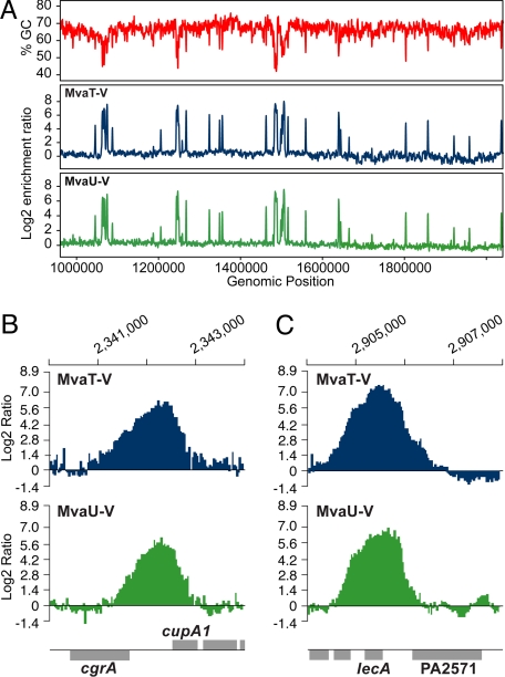
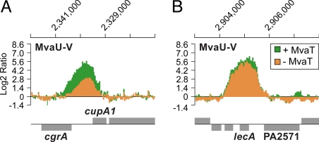
The histone-like nucleoid structuring protein, H-NS, is a prominent global regulator of gene expression. Many Gram-negative bacteria contain multiple members of the H-NS family of proteins. Thus, a key question is whether H-NS family members have overlapping or distinct functions. To address this question we performed genome-wide location analyses with MvaT and MvaU, the two H-NS family members present in Pseudomonas aeruginosa. We show that MvaT and MvaU bind the same chromosomal regions, coregulating the expression of approximately 350 target genes. We show further that like H-NS in enteric bacteria, which functions as a transcriptional silencer of foreign DNA by binding to AT-rich elements, MvaT and MvaU bind preferentially to AT-rich regions of the chromosome. Our findings establish that H-NS paralogs can function coordinately to regulate expression of the same set of target genes, and suggest that MvaT and MvaU are involved in silencing foreign DNA elements in P. aeruginosa.


Conflict of interest statement

The authors declare no conflict of interest.

Figures

Fig. 1.
Fig. 1.
MvaT and MvaU occupy the same AT-rich regions of the chromosome. (A) The percent GC content for 1 Mb of the PAO1 chromosome (in windows of 1000 bp across the genome with a window step size of 500 bp) is plotted (in red) against the locations of MvaT-V (plotted as log2 ratios in blue) and MvaU-V (plotted as log2 ratios in green) as determined by ChIP-on-chip. Association of MvaT-V and MvaU-V with the cgrA-cupA1 intergenic region (B), and with the lecA promoter and coding region (C). Log2 ratio values were normalized and averaged across three replicate arrays.
Fig. 2.
Fig. 2.
MvaT can influence the association of MvaU with target sites. Association of MvaU-V with the cgrA-cupA1 intergenic region (A) and with the lecA promoter and coding region (B), in the presence (green) or absence (orange) of MvaT. Log2 ratio values were normalized and averaged across three replicate arrays.
Fig. 3.
Fig. 3.
Combined loss of MvaT and MvaU results in lethality. (A) ClpXP-based controllable protein degradation system. Representation of the VSVG-DAS4 tag integration vector and its use in making strains synthesizing MvaT-V-DAS4 and MvaU-V-DAS4. The VSV-G epitope tag (blue) and the DAS4 tag (green) are shown together with the gentamicin resistance determinant (GentR), the mobilization region (mob) and FRT sites. (B) Depletion of MvaT-V-DAS4 in a ΔmvaU mutant strain and depletion of MvaU-V-DAS4 in a ΔmvaT mutant strain results in loss of viability. Plasmids pV-SspB and pPSPK-SspB express V-sspB and sspB respectively in an IPTG-inducible fashion. Plasmids pPSV35 and pPSPK are empty vector controls. Colonies of plasmid-containing cells were resuspended in LB to the same OD600. Cells were serially diluted to the indicated OD600 and spotted onto LB plates that either did (+) or did not (−) contain 2 mM IPTG. Row 1, PAO1 ΔsspB cupA lacZ MvaT-V-DAS4 carrying pV-SspB. Row 2, PAO1 ΔsspB ΔmvaU cupA lacZ MvaT-V carrying pV-SspB. Row 3, PAO1 ΔsspB ΔmvaU cupA lacZ MvaT-V-DAS4 carrying pPSV35. Row 4, PAO1 ΔsspB ΔmvaU cupA lacZ MvaT-V-DAS4 carrying pV-SspB. Row 5, PAO1 ΔsspB MvaU-V-DAS4 carrying pPSPK-SspB. Row 6, PAO1 ΔsspB ΔmvaT MvaU-V carrying pPSPK-SspB. Row 7, PAO1 ΔsspB ΔmvaT MvaU-V-DAS4 carrying pPSPK. Row 8, PAO1 ΔsspB ΔmvaT MvaU-V-DAS4 carrying pPSPK-SspB.
Fig. 4.
Fig. 4.
Combined loss of MvaT and MvaU has a larger effect on the expression of target genes than loss of MvaT or MvaU alone. Cells of the MvaT depletion strains containing plasmid pV-SspB (expressing V-SspB in an IPTG-inducible manner) or the empty control vector pPSV35, were first grown to mid-log (time 0), then grown for a further 30 min either in the absence (−) or presence of IPTG (+). Cells were harvested at the indicated time points and analyzed for protein and RNA. (A) Depletion of MvaT-V-DAS4 and induction of V-SspB analyzed by western blot. (Upper) Western blot analysis probed with antibody against VSV-G tag. (Lower) Western blot probed with antibody against the α subunit of RNA polymerase serves as a control for sample loading. PAO1 ΔsspB cupA lacZ MvaT-V-DAS4 carrying either pPSV35 (lanes 1–3) or pV-SspB (lanes 4–6). PAO1 ΔsspB ΔmvaU cupA lacZ MvaT-V-DAS4 carrying either pPSV35 (lanes 7–9) or pV-SspB (lanes 10–12). (B) Effect on target gene expression of depleting MvaT in the absence and presence of MvaU. Abundance of transcripts in cells PAO1 ΔsspB ΔmvaU cupA lacZ MvaT-V-DAS4 (ΔmvaU strain) carrying pV-SspB relative to those carrying pPSV35 (black bars). Abundance of transcripts in cells of PAO1 ΔsspB cupA lacZ MvaT-V-DAS4 (mvaU+ strain) carrying pV-SspB relative to those carrying pPSV35 (gray bars). The indicated transcripts were quantified by qRT-PCR. The dotted line at the bottom of the graph represents transcript abundance in cells of the strains carrying the pPSV35 control vector. (C) Effect of ΔmvaT and ΔmvaU mutations on target gene expression. Abundance of transcripts in cells of PAO1 ΔmvaT cupA lacZ (black bars) and cells of PAO1 ΔmvaU cupA lacZ (gray bars) relative to those in cells of the WT control PAO1 cupA lacZ. The dotted line represents the transcript levels of PAO1 cupA lacZ. Error bars in B and C represent relative expression values calculated from +/−1 SD from the mean ΔΔCt.

References

    1. Dorman CJ. H-NS: A universal regulator for a dynamic genome. Nat Rev Microbiol. 2004;2:391–400. - PubMed
    1. Grainger DC, Hurd D, Goldberg MD, Busby SJW. Association of nucleoid proteins with coding and non-coding segments of the Escherichia coli genome. Nucleic Acids Res. 2006;34:4642–4652. - PMC - PubMed
    1. Lucchini S, et al. H-NS mediates the silencing of laterally acquired genes in bacteria. PLoS Pathog. 2006;2:e81. - PMC - PubMed
    1. Oshima T, Ishikawa S, Kurokowa K, Aiba H, Ogasawara N. Escherichia coli histone-like protein H-NS preferentially binds to horizontally acquired DNA in association with RNA polymerase. DNA Res. 2006;13:141–153. - PubMed
    1. Navarre WW, et al. Selective silencing of foreign DNA with low GC content by the H-NS protein in Salmonella. Science. 2006;313:236–238. - PubMed

Publication types

MeSH terms

LinkOut - more resources