. 2008 Dec 2;105(48):18907-12.
doi: 10.1073/pnas.0810111105. Epub 2008 Nov 21.

Notch and Kras reprogram pancreatic acinar cells to ductal intraepithelial neoplasia

Affiliations

Notch and Kras reprogram pancreatic acinar cells to ductal intraepithelial neoplasia

Jean-Paul De La O et al. Proc Natl Acad Sci U S A. .

Abstract

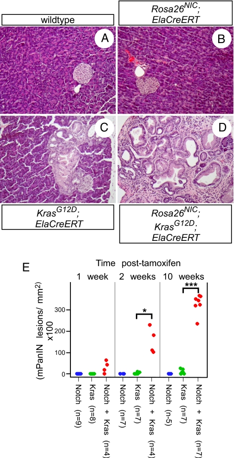
Efforts to model pancreatic cancer in mice have focused on mimicking genetic changes found in the human disease, particularly the activating KRAS mutations that occur in pancreatic tumors and their putative precursors, pancreatic intraepithelial neoplasia (PanIN). Although activated mouse Kras mutations induce PanIN lesions similar to those of human, only a small minority of cells that express mutant Kras go on to form PanINs. The basis for this selective response is unknown, and it is similarly unknown what cell types in the mature pancreas actually contribute to PanINs. One clue comes from the fact that PanINs, unlike most cells in the adult pancreas, exhibit active Notch signaling. We hypothesize that Notch, which inhibits differentiation in the embryonic pancreas, contributes to PanIN formation by abrogating the normal differentiation program of tumor-initiating cells. Through conditional expression in the mouse pancreas, we find dramatic synergy between activated Notch and Kras in inducing PanIN formation. Furthermore, we find that Kras activation in mature acinar cells induces PanIN lesions identical to those seen upon ubiquitous Kras activation, and that Notch promotes both initiation and dysplastic progression of these acinar-derived PanINs, albeit short of invasive adenocarcinoma. At the cellular level, Notch/Kras coactivation promotes rapid reprogramming of acinar cells to a duct-like phenotype, providing an explanation for how a characteristically ductal tumor can arise from nonductal acinar cells.


Conflict of interest statement

The authors declare no conflict of interest.

Figures

Fig. 1.
Fig. 1.
Notch and Kras synergize to induce mPanIN initiation. (A–I) H&E-stained sections from mice of the indicated genotypes, administered TM in utero (A–F) or left untreated (G–I). Broad mosaic activation of Rosa26NIC, via in utero TM treatment, produces foci of convoluted, undifferentiated epithelium (D, bracket), whereas broad activation of KrasG12D produces isolated mPanIN lesions (E, arrow). Broad coactivation of both alleles results in almost complete replacement of normal exocrine tissue by mPanIN-like epithelium (C and F). Focal mosaic activation of NotchIC or Kras alone, in the absence of TM, produces no or few abnormalities (G and H), whereas focal coactivation of both alleles induces numerous mPanIN lesions (I, arrow). Also indicated are normal ducts (du) and islets (is). (Magnification: A–C 100×, D–F 400×, G–I 200×.) (J) Stripchart of mPanIN initiation indices for individual TM-untreated Pdx1CreERT mice, expressing activated Notch1 (Notch) and/or KrasG12D (Kras), at indicated ages. P-values (by t test): *, P < 0.05, **, P < 0.01.
Fig. 2.
Fig. 2.
Acinar-specific activation of KrasG12D induces mPanIN lesions similar to those induced with general pancreatic Cre drivers. (A and B) H&E-staining of mPanIN lesions in KrasG12D;ElaCreERT mice. Both low-grade mPanIN-1 lesions are found, with columnar cytoplasm and basally located nuclei (A), as well as rarer high-grade lesions, including an mPanIN-3 lesion with nuclear atypia and luminal budding (B). (C) PAS staining (magenta) of acinar-derived mPanIN. (D) Cytokeratin-19 staining (brown) of acinar-derived mPanIN. (Magnification: 400×.)
Fig. 3.
Fig. 3.
Notch and Kras synergize to induce mPanINs from acinar cells. (A–D) Representative H&E-stained sections from pancreata of the indicated genotypes, 10 weeks postTM administration. Acinar-specific NotchIC expression has no detectable effect (B), whereas acinar-specific KrasG12D activation induces focal mPanINs (C). Coactivation of the alleles induces many mPanIN lesions, replacing most of the normal exocrine tissue (D). (Magnification: 200×.) (E) Stripchart indicating mPanIN initiation indices of individual ElaCreERT mice, expressing NotchIC and/or KrasG12D, at indicated timepoints following tamoxifen treatment. P-values: *, P < 0.05; ***, P < 0.0005.
Fig. 4.
Fig. 4.
Notch and Kras synergize to induce rapid acinar-to-ductal reprogramming in vivo. Adult (6-week-old) mice of the indicated genotypes were treated with tamoxifen and analyzed two weeks later. (A–L) Immunofluorescence staining for GFP (green, indicating activation of Rosa26NIC), the acinar marker amylase (red) and the duct marker CK19 (white). Activation of Notch in acinar cells results in no detectable changes (E–H, note green nuclei in amylase+ acinar cells, negative for CK19), whereas Notch/Kras coactivation results in ductal reprogramming specifically of GFP-positive cells, as indicated by loss of amylase and up-regulation of CK19 (I–L). (M–X) Immunofluorescence staining for GFP (green), the acinar regulatory transcription factor Ptf1a (red) and CK19 (white) provides further evidence for Notch/Kras-driven reprogramming, as NotchIC expression alone does not result in loss of Ptf1a expression (Q–T), whereas NotchIC/KrasG12D coactivation down-regulates Ptf1a although inducing CK19 (U–X). (Magnification: 400×.)

References

    1. Maitra A, Hruban RH. Pancreatic cancer. Annu Rev Pathol. 2008;3:157–188. - PMC - PubMed
    1. Hingorani SR, et al. Preinvasive and invasive ductal pancreatic cancer and its early detection in the mouse. Cancer Cell. 2003;4:437–450. - PubMed
    1. Aguirre AJ, et al. Activated Kras and Ink4a/Arf deficiency cooperate to produce metastatic pancreatic ductal adenocarcinoma. Gene Dev. 2003;17:3112–3126. - PMC - PubMed
    1. Hruban RH, et al. Pathology of genetically engineered mouse models of pancreatic exocrine cancer: Consensus report and recommendations. Cancer Res. 2006;66:95–106. - PubMed
    1. Jensen J. Gene regulatory factors in pancreatic development. Dev Dyn. 2004;229:176–200. - PubMed

Publication types

MeSH terms