. 2009 Feb;296(2):L220-8.
doi: 10.1152/ajplung.90474.2008. Epub 2008 Nov 21.

Type 5 phosphodiesterase expression is a critical determinant of the endothelial cell angiogenic phenotype

Affiliations

Type 5 phosphodiesterase expression is a critical determinant of the endothelial cell angiogenic phenotype

Bing Zhu et al. Am J Physiol Lung Cell Mol Physiol. 2009 Feb.

Abstract

Type 5 phosphodiesterase (PDE5) inhibitors increase endothelial cell cGMP and promote angiogenesis. However, not all endothelial cell phenotypes express PDE5. Indeed, whereas conduit endothelial cells express PDE5, microvascular endothelial cells do not express this enzyme, and they are rapidly angiogenic. These findings bring into question whether PDE5 activity is a critical determinant of the endothelial cell angiogenic potential. To address this question, human full-length PDE5A1 was stably expressed in pulmonary microvascular endothelial cells. hPDE5A1 expression reduced the basal and atrial natriuretic peptide (ANP)-stimulated cGMP concentrations in these cells. hPDE5A1-expressing cells displayed attenuated network formation on Matrigel in vitro and also produced fewer blood vessels in Matrigel plug assays in vivo; the inhibitory actions of hPDE5A1 were reversed using sildenafil. To examine whether endogenous PDE5 activity suppresses endothelial cell angiogenic potential, small interfering RNA (siRNA) constructs were stably expressed in pulmonary artery endothelial cells. siRNA selectively decreased PDE5 expression and increased basal and ANP-stimulated cGMP concentrations in these conduit cells. PDE5 downregulation increased network formation on Matrigel in vitro and increased blood vessel formation in Matrigel plug assays in vivo. Collectively, our results indicate that PDE5 activity is an essential determinant of angiogenesis and suggest that PDE5 downregulation in microvascular endothelium imparts a stable, enhanced angiogenic potential to this cell type.


Figures

Fig. 1.
Fig. 1.
Overexpression of human type 5 phosphodiesterase (PDE5) A1 gene in pulmonary microvascular endothelial cells (PMVECs) reduces cellular cGMP. A: retroviral construct of human PDE5A1 with EGFP fusion was used to transfect PMVECs. B: Western blot analysis resolved a 125-kDa protein in hPDE5A1/EGFP expressing cells (5A1) using both GFP and human PDE5-specific antibodies (38, 40), which was confirmed by coimmunoprecipitation (IP:GFP and IB: hPDE5). C: the hPDE5A1/EGFP expressing cells possessed a greater than 50-fold increase in cGMP-hydrolytic activity, whereas no change in cAMP-PDE activity was detected (inset). D: the hPDE5A1 enzyme was inhibited by the PDE5 selective inhibitor, sildenafil, IC50 = 20 nM. E: a decrease in basal cGMP was detected in hPDE5A1-expressing cells compared with PMVECs. F: expression of hPDE5A1 in PMVECs attenuated the increase in cGMP after atrial natriuretic peptide (ANP) (10 nM) stimulation compared with previously reported responses in PDE5-deficient PMVECs (39). Pretreatment of hPDE5A1-expressing cells with sildenafil (100 nM for 10 min) increased basal cGMP concentrations and increased the ANP-induced rise in cGMP. *P < 0.05, n = 3 compared with PMVECs or hPDE5A1 controls.
Fig. 2.
Fig. 2.
PDE5 small interfering RNA (siRNA) decreases rat PDE5 mRNA in pulmonary artery endothelial cells (PAECs). A: 6 siRNA sequences (Si-1 to -6) targeting NH2-terminal domains specific to rat (r) and/or human (h) PDE5A1 and PDE5A2 genes were designed using a gene-walking approach. The siRNAs were transfected into PAECs using retroviral constructs for establishing stable cell lines. B: 4 rat-specific siRNAs, siRNA-3, -4, -5, and -6, effectively inhibited both rPDE5A1 and rPDE5A2 mRNA in PAECs. C: inhibition of PDE5A1/5A2 mRNA was greatest in siRNA-3-expressing cells and was confirmed by RQ RT-PCR using 18S and PDE4D5 (4D5) as the endogenous controls (10). siRNAs consistently decreased rPDE5A1 protein levels (D) and enzyme activities (E). siRNA-3 inhibited more than 90% of PDE5 activity. Negative controls, scrambled RNA controls, and siRNA-1 specific to human PDE5A1 did not change rPDE5A1 and rPDE5A2 at mRNA levels (B), protein levels (D), or activity (E). None of the siRNA constructs changed expression of endogenous cAMP-PDE isoforms, including PDE4D5 (4D5, B and C) and PDE4A (4A, D), and did not influence cAMP-PDE activity in PMVECs (E). *P < 0.05, n = 3 compared with controls.
Fig. 3.
Fig. 3.
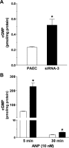
Inhibition of PDE5 expression in PAECs increases cGMP. Inhibition of PDE5 expression in PAECs (open bars) by siRNA-3 (closed bars) potentiates cGMP levels under basal (A) and ANP-stimulated (10 nM) conditions (B). *P < 0.05, n = 3 compared with PAECs control.
Fig. 4.
Fig. 4.
PDE5 regulates endothelial cell growth. A and C: hPDE5A1-expressing PMVECs and wild-type PMVECs showed similar growth under 10% serum conditions, whereas siRNA-3-expressing cells displayed a greater proliferative response than did PAECs. B and D: under 0.5% serum culture conditions, hPDE5A1-expressing cell showed a slower growth than PMVECs. Under the same low-serum culture, siRNA-3 cells showed a greater increase in cell number than did PAECs. Cells were cultured in 60-mm dishes for cell counting, n = 4. Note the different y-axes scales (A and C, 10% serum; B and D, 0.5% serum). *P < 0.05 compared with controls.
Fig. 5.
Fig. 5.
Overexpression of hPDE5A1 in PMVECs inhibits network formation on Matrigel and decreases angiogenesis. A: reduced network formation was seen in hPDE5A1-expressing cells compared with PMVEC controls, which was reversed when cells were pretreated with sildenafil (100 nM) for 24 h. Branch-like tubes, or microspikes, were seen in hPDE5A1 cells after 24–48 h in culture, which were also eliminated by sildenafil pretreatment. B: network formation of PMVECs (closed squares) on Matrigel was counted over the time of cell culture. Overexpression of hPDE5A1 delayed network formation and reduced the total number of networks. The decrease in network formation observed in hPDE5A1-expressing cells (open squares) was acutely reversed by sildenafil pretreatment (open circles). 2 × 10³ cells/well were seeded on Matrigel in 96-well plates, n = 4. *P < 0.05 compared with control PMVECs. C: dynamic changes of branch-like tubes, or microspikes, grown on Matrigel were seen in hPDE5A1-expressing cells. Solid arrows indicate those tubes undergoing a growth and elongation response, whereas dashed arrows indicate tubes undergoing regression. *Center of the network.
Fig. 6.
Fig. 6.
Inhibition of PDE5 expression in PAECs promotes network formation on Matrigel. A: inhibition of PDE5 activity using siRNA-3 increases network formation on Matrigel culture when compared with wild-type PAECs. B: numbers of networks counted on Matrigel indicated more networks in siRNA-3-expressing cells than in PAECs. 5 × 10⁴ cells/well were seeded on Matrigel in 96-well plates, n = 4. C: pretreatment of PAECs with PDE5-specific inhibitor sildenafil (100 nM, 24 h) increased network formation on Matrigel. Inset: PAECs network formation with sildenafil pretreatment after 72 h of culture on Matrigel. *P < 0.05 compared with controls.
Fig. 7.
Fig. 7.
PDE5 regulates vessel formation in Matrigel plug assay in vivo. A: hPDE5A1-expressing cells possessed fewer vessels in Matrigel plugs. B: siRNA-3 cells showed more vessel formation in Matrigel plugs. Each animal was injected with Matrigel-cell mixtures, and plugs were harvested after 10 days for vessel staining assay. N = 4, *P < 0.05 compared with PMVEC or PAEC controls.

References

    1. Al-Ani B, Hewett PW, Ahmed S, Cudmore M, Fujisawa T, Ahmad S, Ahmed A. The release of nitric oxide from S-nitrosothiols promotes angiogenesis. PLoS ONE 1: e25, 2006. - PMC - PubMed
    1. Alvarez DF, Huang L, King JA, Elzarrad MK, Yoder MC, Stevens T. Lung microvascular endothelium is enriched with progenitor cells that exhibit vasculogenic capacity. Am J Physiol Lung Cell Mol Physiol 294: L419–L430, 2008. - PubMed
    1. Benedict N, Seybert A, Mathier MA. Evidence-based pharmacologic management of pulmonary arterial hypertension. Clin Ther 29: 2134–2153, 2007. - PubMed
    1. Bussolati B, Dunk C, Grohman M, Kontos CD, Mason J, Ahmed A. Vascular endothelial growth factor receptor-1 modulates vascular endothelial growth factor-mediated angiogenesis via nitric oxide. Am J Pathol 159: 993–1008, 2001. - PMC - PubMed
    1. Carmeliet P Angiogenesis in life, disease and medicine. Nature 438: 932–936, 2005. - PubMed

Publication types

MeSH terms

LinkOut - more resources